
Register now and get the free poster
Steel Quality Rating: Free poster
Steel suppliers and industry manufacturers face the challenge of being compliant with multiple ever-stricter standards. These different standards classify non-metallic inclusions to determine the quality of steel.
From this poster, you get an overview of the following key points related to the rating of non-metallic inclusions:
- International and regional standards
- Classification of individual particles (dimension, shape, aspect ratio, color, and grouping)
- Comparison of inclusion types defined in the most common standards
- Classification of severity of the inclusions
The Steel Quality Solution Suite provides several complete workflow solutions to rate non-metallic inclusions rapidly and reliably.
- Detection and classification of non-metallic inclusions by manual and automated image analysis
- Easy guidance through data acquisition and analysis
- Fast and easy review of rated inclusions with a simultaneous sample overview and detailed view of regions of interest
- Quick adaptation to changes in international, regional, and organizational rating standards

Related Articles

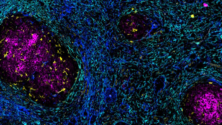
IBEX, Cell DIVE, and RNA-Seq: A Multi-omics Approach to Follicular Lymphoma
In a recent study by Radtke et al., a multi-omics spatial biology approach helps shed light on early relapsing lymphoma patients

Overcoming Observational Challenges in Organoid 3D Cell Culture
Learn how to overcome challenges in observing organoid growth. Read this article and discover new solutions for real-time monitoring which do not disturb the 3D structure of the organoids over time.
 at the edge of a battery electrode acquired with a DVM6 digital microscope.")
Burr Detection During Battery Manufacturing
See how optical microscopy can be used for burr detection on battery electrodes and determination of damage potential to achieve rapid and reliable quality control during battery manufacturing.

How do Cells Talk to Each Other During Neurodevelopment?
Professor Silvia Capello presents her group’s research on cellular crosstalk in neurodevelopmental disorders, using models such as cerebral organoids and assembloids.

Battery Particle Detection During the Production Process
How battery particle detection and analysis is enhanced with optical microscopy and laser spectroscopy for rapid, reliable, and cost-effective QC during battery production is explained in this…
 where cyan indicates nuclei and magenta tight junctions.")
Rapid Check of Live Stem Cells in Cell-Culture Inserts set in Multi-Well Plates
See how efficient imaging of live iPSC stem cells within cell-culture inserts set in a multi-well plate can be done to evaluate the cells using a THUNDER Imager. Just read this article.

Automatic Alignment of Sample and Knife for High Sectioning Quality
Automatic alignment of sample and knife on the ultramicrotome UC Enuity, enabling even untrained users to create ultrathin sections with reduced risk of losing precious sections.

High Quality Sectioning in Ultramicrotomy
Discover the significance of achieving high-quality uniform sections with ultramicrotomy for precise imaging in electron microscopy.

Extended Live-cell Imaging at Nanoscale Resolution
Extended live-cell imaging with TauSTED Xtend. Combined spatial and lifetime information allow super-resolution microscopy at extremely low light dose.
, actin network (ATTO 647N), and nuclear pore basket (CF 680R).")
The Guide to STED Sample Preparation
This guide is intended to help users optimize sample preparation for stimulated emission depletion (STED) nanoscopy, specifically when using the STED microscope from Leica Microsystems. It gives an…

How to Streamline Your Histology Workflows
Streamline your histology workflows. The unique Fluosync detection method embedded into Mica enables high-res RGB color imaging in one shot.
 dataset, showing the biochemically distinct structures of a fresh, untreated apple slice.")
How to Prepare Samples for Stimulated Raman Scattering (SRS) imaging
Find here guidelines for how to prepare samples for stimulated Raman scattering (SRS), acquire images, analyze data, and develop suitable workflows. SRS spectroscopic imaging is also known as SRS…
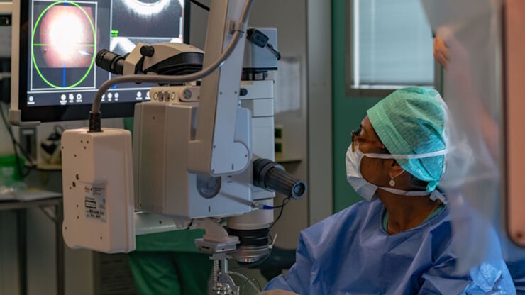
RPE65 Gene Therapy Subretinal Injection: Benefits of Intraoperative OCT
Discover how RPE65 gene therapy subretinal injection procedures in patients with Leber congenital amaurosis is supported by intraoperative Optical Coherence Tomography.
Accelerating Discovery for Multiplexed Imaging of Diverse Tissues
Explore IBEX: Open-source multiplexed imaging. Join the collaborative IBEX Imaging Community for optimized tissue processing, antibody selection, and human atlas construction.

Key Factors for Efficient Cleanliness Analysis
An overview of the key factors necessary for technical cleanliness and efficient cleanliness analysis concerning automotive and electronics manufacturing and production is provided in this article.

Technical Terms for Digital Microscope Cameras and Image Analysis
Learn more about the basic principles behind digital microscope camera technologies, how digital cameras work, and take advantage of a reference list of technical terms from this article.
 and oblique (right) brightfield illumination using a Leica compound microscope. The defect on the wafer surface is clearly more visible with oblique illumination.")
Rapid Semiconductor Inspection with Microscope Contrast Methods
Semiconductor inspection for QC of materials like wafers can be challenging. Microscope solutions that offer several contrast methods enable fast and reliable defect detection and efficient workflows.

Transforming Multiplexed 2D Data into Spatial Insights Guided by AI
Aivia 13 handles large 2D images and enables researchers to obtain deep insights into microenvironment surrounding their phenotypes with millions of detected objects and automatic clustering up to 30…
Related Articles
IBEX, Cell DIVE, and RNA-Seq: A Multi-omics Approach to Follicular Lymphoma
In a recent study by Radtke et al., a multi-omics spatial biology approach helps shed light on early relapsing lymphoma patients
Overcoming Observational Challenges in Organoid 3D Cell Culture
Learn how to overcome challenges in observing organoid growth. Read this article and discover new solutions for real-time monitoring which do not disturb the 3D structure of the organoids over time.
Burr Detection During Battery Manufacturing
See how optical microscopy can be used for burr detection on battery electrodes and determination of damage potential to achieve rapid and reliable quality control during battery manufacturing.
How do Cells Talk to Each Other During Neurodevelopment?
Professor Silvia Capello presents her group’s research on cellular crosstalk in neurodevelopmental disorders, using models such as cerebral organoids and assembloids.
Battery Particle Detection During the Production Process
How battery particle detection and analysis is enhanced with optical microscopy and laser spectroscopy for rapid, reliable, and cost-effective QC during battery production is explained in this…
Rapid Check of Live Stem Cells in Cell-Culture Inserts set in Multi-Well Plates
See how efficient imaging of live iPSC stem cells within cell-culture inserts set in a multi-well plate can be done to evaluate the cells using a THUNDER Imager. Just read this article.
Automatic Alignment of Sample and Knife for High Sectioning Quality
Automatic alignment of sample and knife on the ultramicrotome UC Enuity, enabling even untrained users to create ultrathin sections with reduced risk of losing precious sections.
High Quality Sectioning in Ultramicrotomy
Discover the significance of achieving high-quality uniform sections with ultramicrotomy for precise imaging in electron microscopy.
Extended Live-cell Imaging at Nanoscale Resolution
Extended live-cell imaging with TauSTED Xtend. Combined spatial and lifetime information allow super-resolution microscopy at extremely low light dose.
The Guide to STED Sample Preparation
This guide is intended to help users optimize sample preparation for stimulated emission depletion (STED) nanoscopy, specifically when using the STED microscope from Leica Microsystems. It gives an…
How to Streamline Your Histology Workflows
Streamline your histology workflows. The unique Fluosync detection method embedded into Mica enables high-res RGB color imaging in one shot.
How to Prepare Samples for Stimulated Raman Scattering (SRS) imaging
Find here guidelines for how to prepare samples for stimulated Raman scattering (SRS), acquire images, analyze data, and develop suitable workflows. SRS spectroscopic imaging is also known as SRS…
RPE65 Gene Therapy Subretinal Injection: Benefits of Intraoperative OCT
Discover how RPE65 gene therapy subretinal injection procedures in patients with Leber congenital amaurosis is supported by intraoperative Optical Coherence Tomography.
Accelerating Discovery for Multiplexed Imaging of Diverse Tissues
Explore IBEX: Open-source multiplexed imaging. Join the collaborative IBEX Imaging Community for optimized tissue processing, antibody selection, and human atlas construction.
Key Factors for Efficient Cleanliness Analysis
An overview of the key factors necessary for technical cleanliness and efficient cleanliness analysis concerning automotive and electronics manufacturing and production is provided in this article.
Technical Terms for Digital Microscope Cameras and Image Analysis
Learn more about the basic principles behind digital microscope camera technologies, how digital cameras work, and take advantage of a reference list of technical terms from this article.
Rapid Semiconductor Inspection with Microscope Contrast Methods
Semiconductor inspection for QC of materials like wafers can be challenging. Microscope solutions that offer several contrast methods enable fast and reliable defect detection and efficient workflows.
Transforming Multiplexed 2D Data into Spatial Insights Guided by AI
Aivia 13 handles large 2D images and enables researchers to obtain deep insights into microenvironment surrounding their phenotypes with millions of detected objects and automatic clustering up to 30…