
Your benefits
- Clarity and accuracy for segmentation
- More time, more data, more images
- AI and application expertise, all in one place


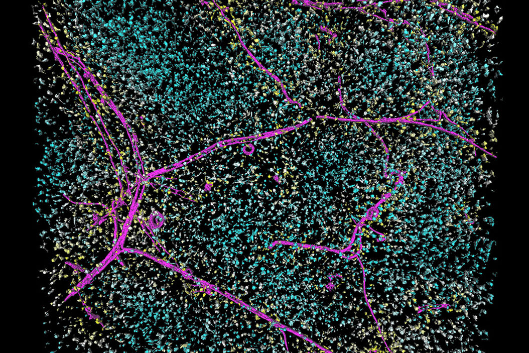
Reach discovery goals with greater accuracy. Crisp THUNDERed images remove the blur and haze you get through conventional widefield imagers, allowing clear marking of signal and background. The powerful synergy of THUNDER and Aivia analyze fluorescen
Increase scientific capabilities
The unique workflow is especially powerful when using thick samples, working with living specimens when low light dosages for excitation are crucial, performing time-lapse experiments, or capturing large image stacks, making it easier for you to more precisely analyze objects of interest. Your dedicated Leica expert has the know-how to get the detailed image acquisition and analysis results you want and need, giving you the freedom to focus on your discoveries.
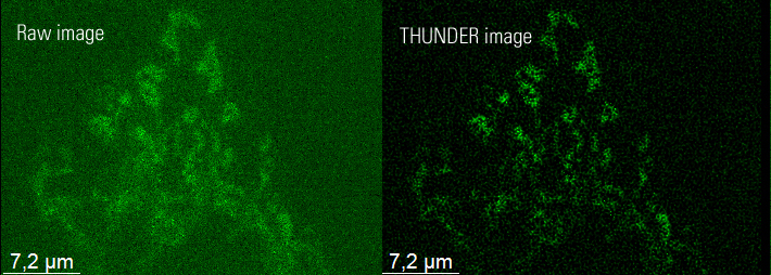
Analysis of THUNDER images
Time lapse showing mitochondria
Compared to the analysis of raw data, a larger number of objects can be seen, indicating a better (more accurate) segmentation. Consequently, the tracking is more precise and any information extracted (displacement, speed, form factor, size, shape) is less prone to error.
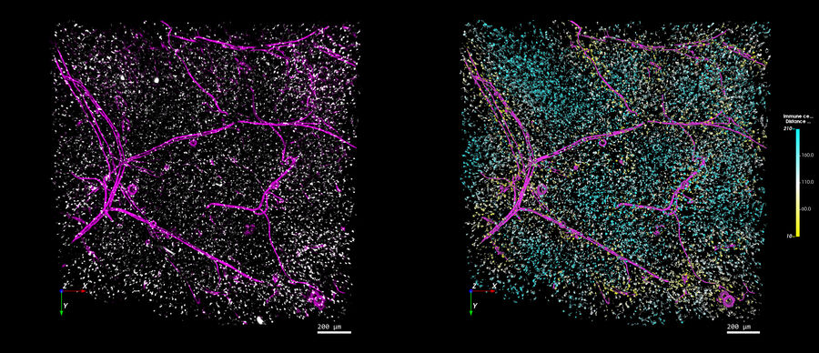
More analysis possibilities with THUNDER and Aivia
On the left you see a THUNDERed image (pink = blood vessel, white = immune cells). On the right there are the rendered surfaces of the vessels colored in pink and the individual immune cells color-coded with their closest distance to the vessels. The legend provides the distance values.
Related Articles
-

Fast, High Acuity Imaging and AI-assisted Analysis
The use of state-of-the-art AI systems is pushing image analysis into a new generation. Challenges…
Jun 01, 2022Read article -

Create New Options for Live Cell Imaging
The use of state-of-the-art AI systems is pushing image analysis into a new generation. Challenges…
Apr 08, 2022Read article -

Accurately Analyze Fluorescent Widefield Images
The specificity of fluorescence microscopy allows researchers to accurately observe and analyze…
Jan 17, 2022Read article
Related Pages
-
THUNDER Imaging Systems
To answer important scientific questions, THUNDER Imaging Systems enable you to obtain a clear view…
Visit related page
