Introduction
Steel is one of the most widely used materials in the world [1]. It is used in a large number of industrial applications, like manufacturing of vehicles, machines, pipelines, and cable towers as well as constructing ships and buildings. Steel quality is critical for these applications, and the rating of non-metallic inclusions (NMIs) in steel is exploited to determine their influence on the quality [2-4]. Fast, precise, and flexible NMI rating solutions which deliver reliable and consistent results offer significant advantages for steel producers and manufacturers of components and parts made with steel. The basic working principles of an automated solution and its advantages for rating steel inclusions are described in this article.
How an Automated NMI Rating Solutions Works
The rating of NMIs to determine steel quality in an efficient and cost-effective way for steel production and industry manufacturing can be achieved, overcoming the challenges arising with manual rating solutions [5], using an automated rating solution.
An automated solution consists of an optical microscope with a motorized stage and objective nosepiece operated with a sophisticated software. It enables a fully automated inclusion rating over the whole steel sample. The typical workflow consists of [2]:
- Definition of the sample and scan area.
- Definition of 5 or 9 focus points in the scan area for data acquisition.
Automatic analysis of the entire sample without the need for users to be present during
Advantages of an Automated NMI Rating Solution
An automated solution offers several advantages when compared to manual NMI rating:
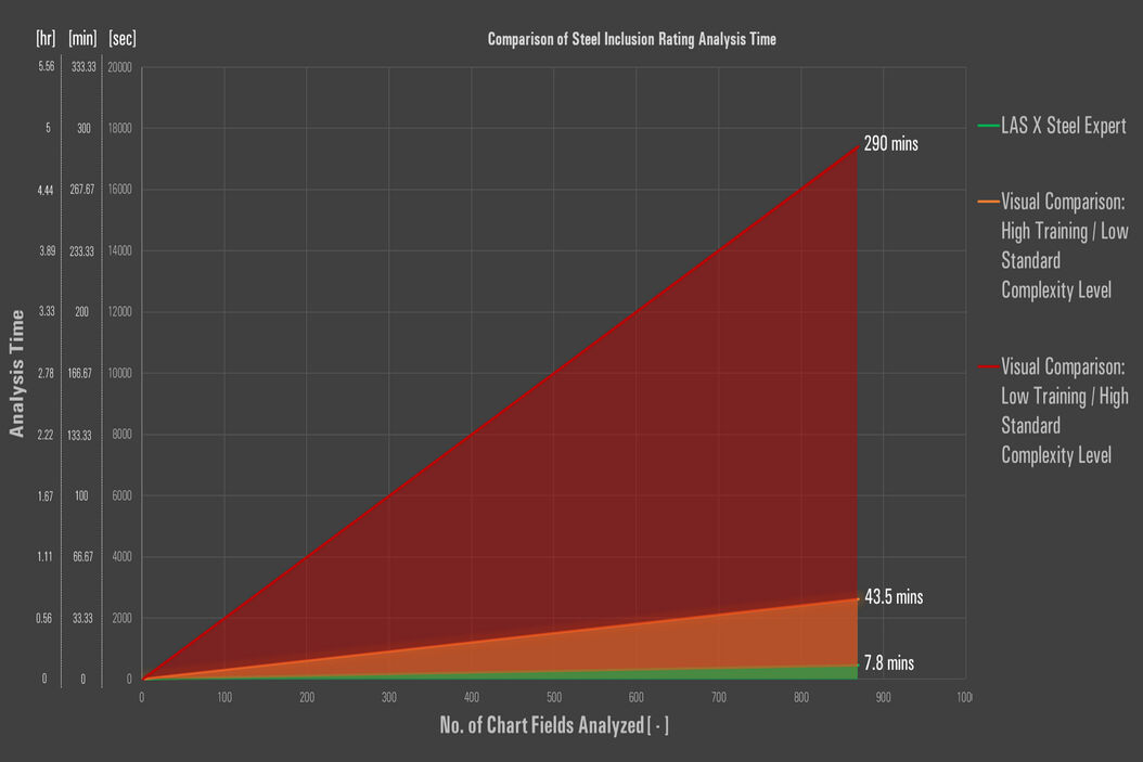
- With careful sample preparation, automated rating enables a fast and reliable analysis of multiple, whole samples
- The automated solution delivers consistent results even if several users are involved or if the system is left unattended during operation
- Advanced review capabilities enable discernment between different types of inclusions or between inclusions and artifacts
- The results obtained are unbiased (independent of the user) and NMI rating is compliant with international, regional, and organizational standards [2,6]
- The software makes all chosen standards available for comparison of the results during a single scan. Inclusion analysis of the same sample can also be done using multiple standards.
An example of an automated NMI rating solution [2] is the Steel Quality Solution Suite from Leica Microsystems utilizing the LAS X Steel Expert software.
References
- J. DeRose, D. Barbero, Reasons Why There is Growing Need for Fast and Reliable Steel Quality Rating Solutions, Science Lab (2020) Leica Microsystems. D. Diez, J. DeRose, T. Locherer, Rate the Quality of Your Steel: Free Webinar and Report: Overview of standard analysis methods and practical solutions for evaluating steel inclusions, Science Lab (2020) Leica Microsystems.
- A. Schué, Steel - It All Depends on What's Really Inside: New European Standard in Steel Industry, Science Lab (2009) Leica Microsystems.
- N. Ecke, Analyzing non-metallic inclusions in steel, Science Lab (2020) Leica Microsystems.
- J. DeRose, D. Barbero, Challenges Faced When Manually Rating Non-Metallic Inclusions (NMIs) to Determine Steel Quality, Science Lab (2020) Leica Microsystems.
- J. DeRose, D. Barbero, T. Locherer, Top Issues Related to Standards for Rating Non-Metallic Inclusions in Steel, Science Lab (2020) Leica Microsystems.
相关文章
-
![[Translate to chinese:] THY1-EGFP labeled neurons in mouse brain processed using the PEGASOS 2 tissue clearing method. Neurons were traced using Aivia’s 3D Neuron Analysis – FL recipe. Image credit: Hu Zhao, Chinese Institute for Brain Research. [Translate to chinese:] THY1-EGFP labeled neurons in mouse brain processed using the PEGASOS 2 tissue clearing method, imaged on a Leica confocal microscope. Neurons were traced using Aivia’s 3D Neuron Analysis – FL recipe. Image credit: Hu Zhao, Chinese Institute for Brain Research.](/fileadmin/_processed_/f/1/csm_Neurons_in_mouse_brain_with_Aivia_3D_Neuron_Analysis_976f72c643.jpg)
借助人工智能,揭示复杂而密集的神经元图像中的洞察
神经元的3D形态学分析通常需要使用不同的成像模式,捕捉多种类型的神经元,并在各种密度下相连的传统Leica SP8显微镜采集多达解神经元的形态,这对许多研究人员来说仍然是一个耗时的挑战。
Jun 16, 2023Read article -

人工智能显微成像能够高效检测稀有事件
对稀有事件进行定位和选择性成像是许多生物样本研究过程的关键。然而,由于时间限制和高度的复杂性,有些实验无法做到,从而限制了获得新发现的前景。通过基于人工智能的显微成像检测稀有事件,这种工作流程将智能样…
Jun 06, 2023Read article -

精确分析宽视野荧光图像
利用荧光显微镜的特异性,即便是使用厚样品和大尺寸样品,研究人员也能够快速轻松地准确观察和分析生物学过程和结构。然而,离焦荧光会提高背景荧光,降低对比度,影响图像的精确分割。THUNDER 与Aivia…
Jan 17, 2022Read article

![[Translate to chinese:] Definition of the sample scan area in the LAS X Steel Expert software.](/fileadmin/_processed_/4/9/csm_Definition_of_the_sample_scan_area_in_the_LAS_X_Steel_Expert_software_18a0bff6f4.png)
![[Translate to chinese:] DMi8 A Inverted Microscope - Professional Configuration](/fileadmin/_processed_/2/5/csm_Inverted_-_NMIR_Professional_Solution_Suite_-_DMi8_A_6837d067a4.jpg)