Clinical Microscope Solutions
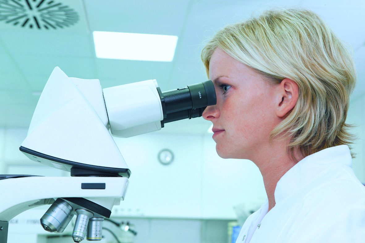
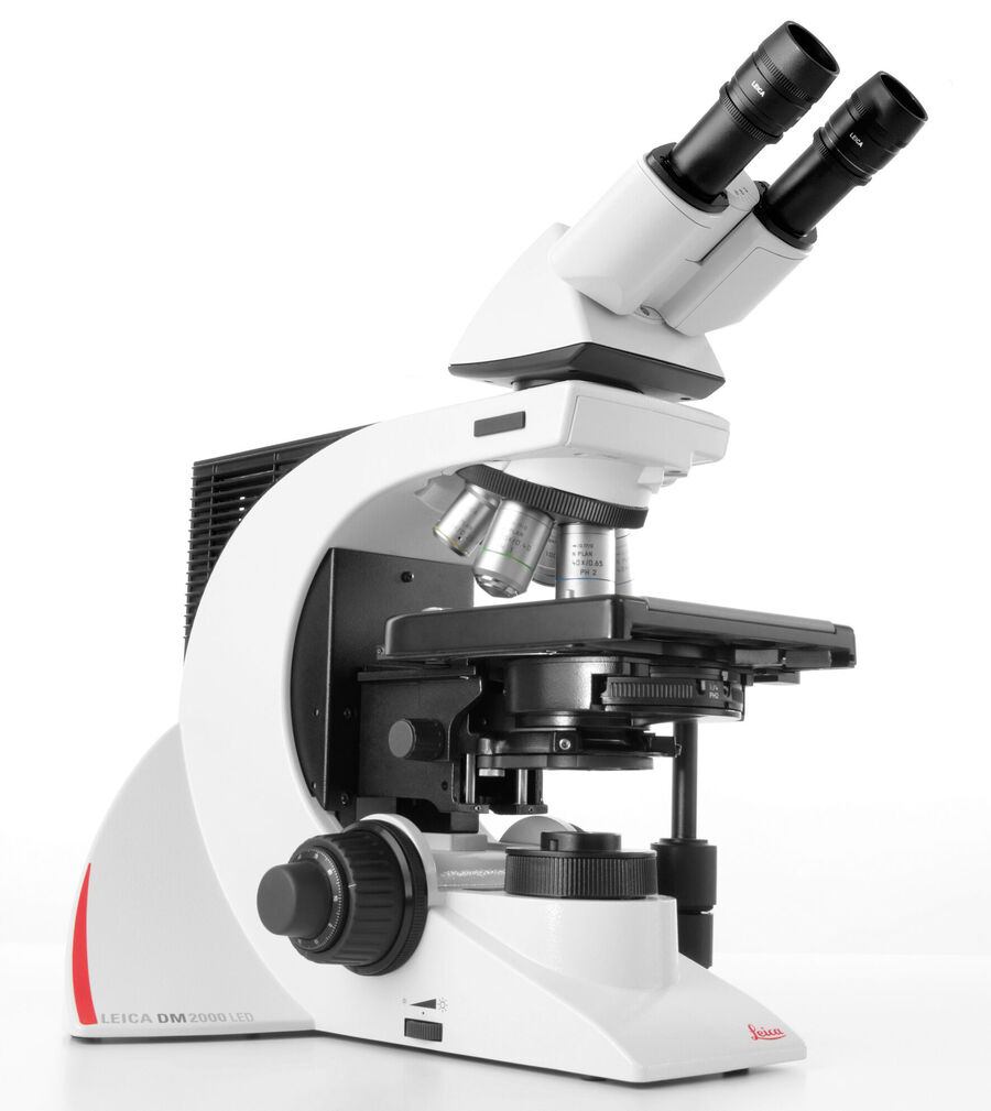
Accuracy and timely diagnoses are crucial in clinical microscopy. Leica clinical microscopes help you diagnose efficiently and accurately as they are equipped with ergonomic accessories, high-quality optics, and true-color cameras.
- Comfortable posture, optimal hand positioning, and less risk of strain: Symmetrical layout of the adjustable ergonomic stage and focus controls
- Reduced risk of eyestrain: Balanced light intensity
- More efficient workflow: Minimal repetitive motions and fast objective change with toggle mode
Customer experiences with a DM3000 microscope for clinical applications
Trudi de Jong and Marianne Noordanus, both from Rotterdam, describe how a DM3000 microscope helps them to perform their clinical microscope work more comfortably and efficiently.
Courtesy of:
Trudi de Jong, Erasmus MC academic hospital Rotterdam, the Netherlands, Hematology
Marianne Noordanus, Star-MDC, Medisch Diagnostisch Centrum, Rotterdam (The Netherlands) Microbiology
Areas of Expertise
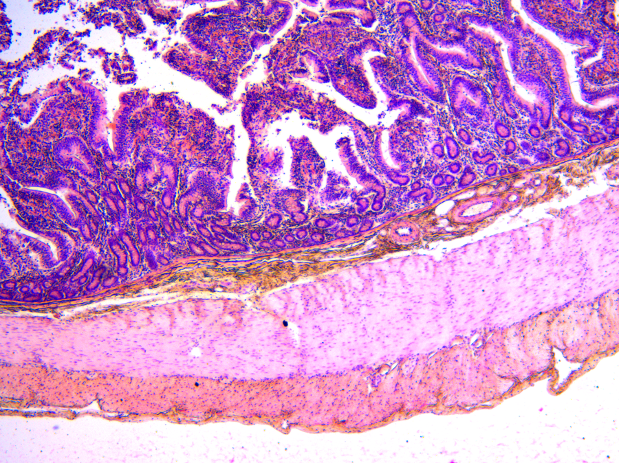
Histopathology:
Image of a H&E-stained duodenum showing the boundary between the submucosal and mucosal regions, making it easy to distinguish the different cell types.
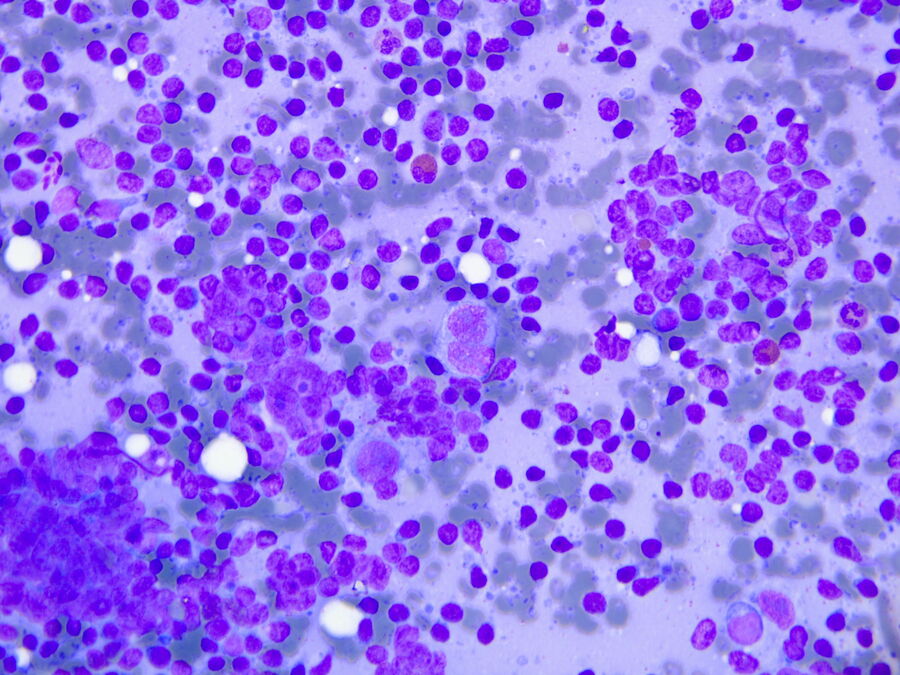
Cytopathology:
Image of tissue stained with Papanicolaou showing hodgkin lymphoma.
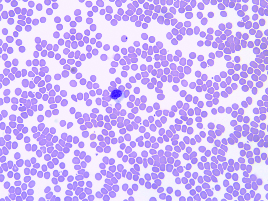
Hematology:
Image of a blood smear used to determine the shape, size, and coloration of red blood cells and the proportions of different white blood cells to see if a patient has any abnormalities.
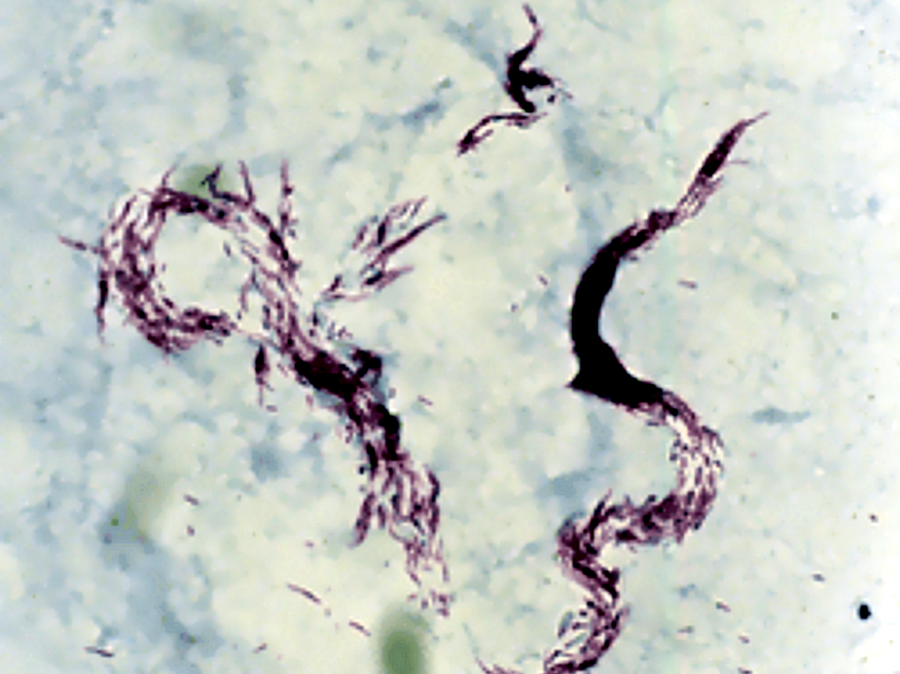
Microbiology:
Image of an acid-fast stained mycobacterium causing tuberculosis. Microbiological examination helps determine if there is an infection caused by microorganisms.
Find the solution for your needs
You can choose from various complete workflow solutions that help you work comfortably and ensure efficient and fast diagnoses.
- Standard: Ideal when performing an occasional diagnosis of specimens where viewing over a selected portion of the slide is necessary.
- Advanced: Ideal for a daily, steady workload performing diagnosis of specimens which are viewed over most of the slide. Also suitable for simultaneous viewing by multiple observers (multiple viewing system) due to its high intensity illumination.
- Professional: Ideal for a high workload performing diagnosis of specimens which require high resolution of detail and viewing over the complete slide. Also suitable for image documentation - can be equipped with a camera.
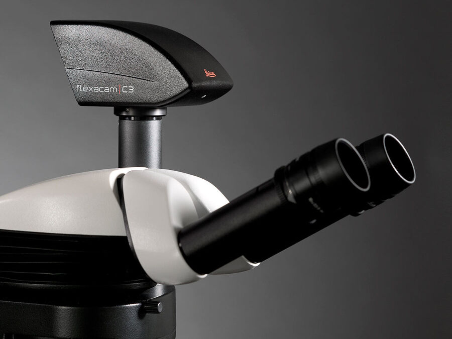
FLEXACAM C3
Easily Document with the 12 MP stand-alone microscope camera
___
Save time and effort with the FLEXACAM C3 microscope camera when capturing, documenting, and sharing images. The camera turns your microscope into a stand-alone digital imaging station with no need for a PC. Start working by simply connecting the FLEXACAM C3 to your microscope, preferred viewing device, and network.
- Capture, document, and share images in seconds
- Stand-alone operation eliminates the need for a PC – only a monitor, or other viewing device, is needed
- Image quality: See more details with crisper images and true-to-life colors
- 12 MP CMOS sensor: Reveals fine details in acquired images
- Fast auto exposure: Allows quick screening of samples with bright and dark areas