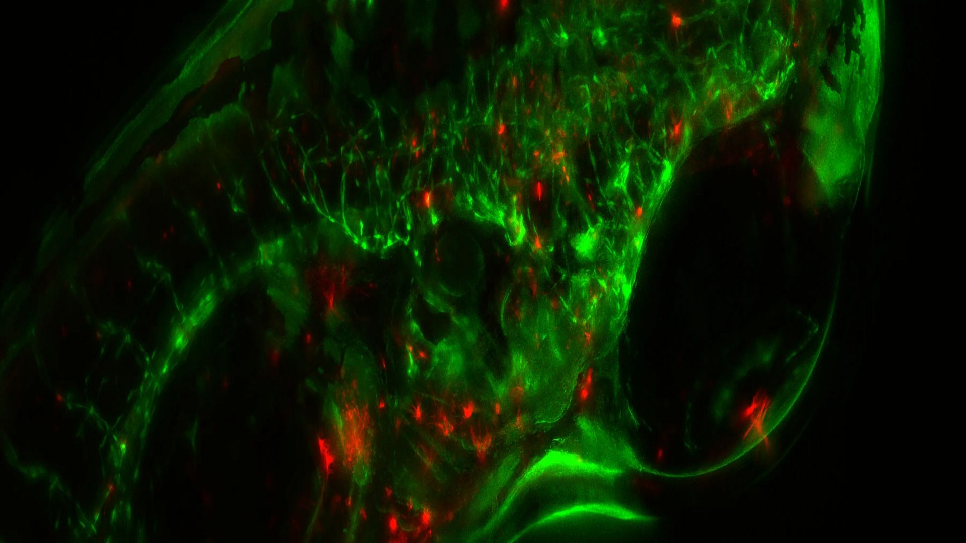
Image of zebrafish embryo at 6 days post fertilization (dpf) showing stable mCherry-expression in hematopoietic progenitor cells (HPCs) and GFP-expressing vascular endothelial cadherin (VEC). Courtesy of Y. Xue, Minnan Normal University, China.
Modular option for superb flexibility
Discover the power of fluorescence combined with flexibility thanks to a modular design. The MZ10 F fluorescence stereo microscope is the ideal solution for applications like gene-expression studies in developmental biology.
- Flexible solution: Change quickly between TripleBeam technology or custom filter sets.
- Modular concept: A wide variety of objectives and accessories to suit your needs.
- Intelligent automation: The LAS X software makes it easy for users to achieve multiple fluorescence recordings.
Fluorescent stereo microscope
See all the details that matter with the M165 FC fully apochromatic corrected stereo microscope.
- Resolve structures down to 1.1 µm in size for detailed fluorescent imaging with the 16.5:1 zoom optics.
- Quick, easy documentation with encoding enabling convenient, reproducible settings.
- Stereo and macro view for manipulation and parallax-free documentation with one system thanks to FluoCombi.
Automated and semi-automated solutions
Capture high-quality images fast with the M205 FCA & M205 FA fluorescence stereo microscopes. Detect fluorescent protein expressions in the earliest stages of transgenic organism development, allowing you to select the right specimen to base your studies on.
- Bright fluorescence signals for a fully illuminated field of view at any magnification.
- See fine details in 3D with higher resolution and more depth of field due to the FusionOptics technology.
- Quick, easy documentation with encoding allowing convenient, reproducible settings.


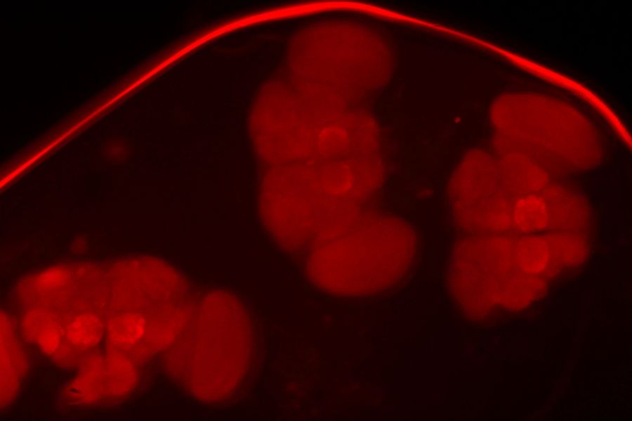
Neurofilaments stained in red to assess neuronal outgrowth in an E12-14 mouse. The mouse was uncleared. Courtesy of Y. Lutz, Centre d’imagerie, IGBMC, France.
Decode 3D biology in real time*
The THUNDER Imager Model Organism allows fast and easy 3D exploration of whole organisms for molecular or developmental biology research.
- Acquire rapidly blur-free images with fine details.
- Keep large model organisms under proper physiological conditions during imaging.
- Simplify your organism handling for an efficient analysis and imaging workflow.
