Workflow Solution for Cryo-Electron Tomography
Cryo-electron tomography (CryoET) is used to resolve biomolecules within their cellular environment down to an unprecedented resolution in the subnanometer range, opening a window to understanding molecular sociology. It is a scale where individual proteins can be identified by their shape alone, without any labeling. But imaging at subnanometer resolution comes with a major challenge: you need to locate and precisely target your sites of interest.
Advanced cryoET solutions from Leica Microsystems use cryo light microscopy for fast, high-resolution imaging and precise transfer of image data, all while maintaining optimal cryo conditions throughout the workflow.
Leica has a unique workflow solution for correlated cryoET. It is called Coral Cryo, which features our STELLARIS, confocal, technology. Read below to learn more about.
Introducing Coral Cryo: the CryoET workflow
Leica's dedicated 3D cryo-electron tomography workflow solutions overcome typical challenges by ensuring sample viability, quality checks and most of all, a precise and reliable 3D targeting mechanism. Make use of our optimized hardware, including cryostage and shuttle, in conjunction with state-of-the-art CryoET targeting software, as well as a variety of seamless integration and transfer options to cryo FIB or VCT stages.
Reduce handling steps between STELLARIS cryo confocal and cryo-FIB
The dedicated Coral Cryo cryo-electron tomography workflow ensures sample viability, quality checks, and most of all, a precise and reliable 3D imaging.
STELLARIS Cryo, including cryo stage and shuttle, is designed to be used in conjunction with LAS X Coral Cryo software, as well as a variety of seamless integration and transfer options to cryo FIB or VCT stages. New cryo stage increases working range in non-cryo applications, expanding STELLARIS usability for confocal imaging.
You can keep the sample safe during the transfer as all components are compatible.

Differentiate targets from background with advanced imaging
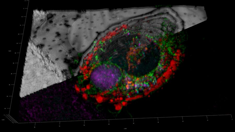
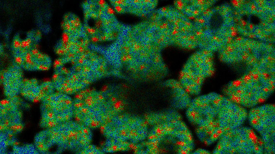
The perfect synergy between our Power HyD detectors family, fully optimized beam path, and next-generation White Light Lasers delivers exceptional imaging performance. Your results are clearer, with greater detail derived from brighter signals, more contrast, and increased sensitivity – even when imaging low-abundance labels and events.
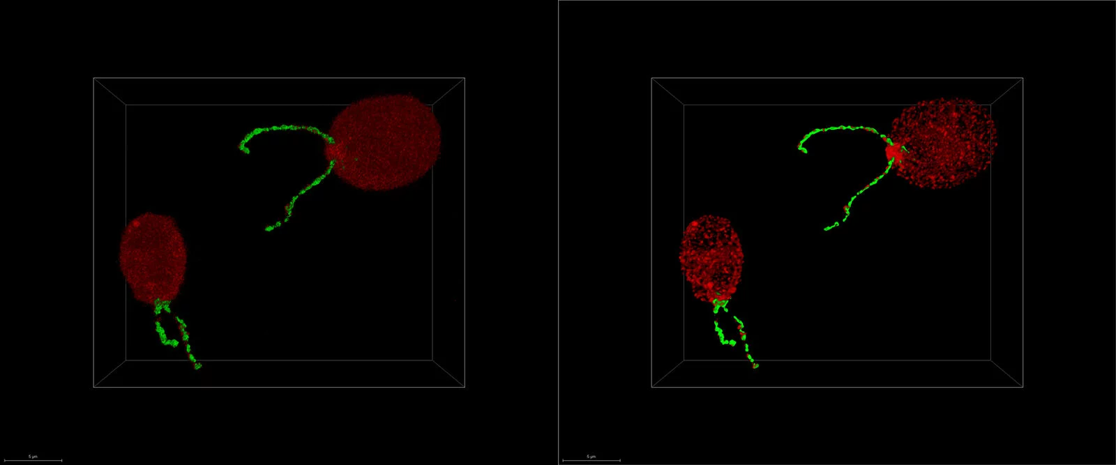
Maximize the information you extract from your vitrified specimen and get in-depth answers to scientific questions with the unique LIGHTNING detection concept, an adaptive extraction process that reveals fine structures and details in the image information.
This allows you to target 3D structures of interest more precisely: in room temperature and under cryogenic conditions, preparing for the next steps in your EM microscopy workflow.