Save up to 40%!
Experience the latest in Ophthalmic Surgery Microscopes
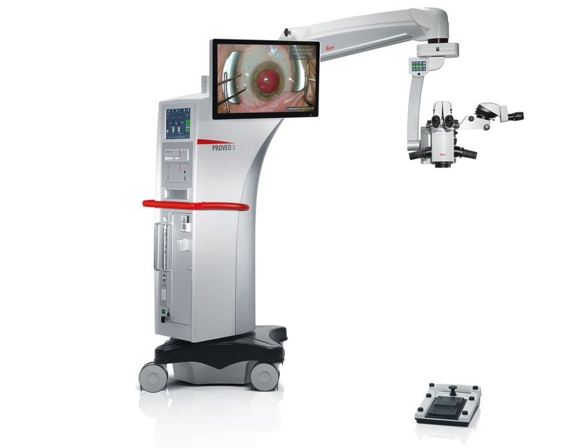
See why the Proveo 8 is Leica’s premier ophthalmic platform for anterior and posterior segment surgery in one microscope. Proveo 8 provides solutions for the most complex procedures as well as being a powerhouse for routine ophthalmic surgery.
Have Questions? Leica's team of product specialists are available to answer with solutions to meet any level of ophthalmic surgery need.
Contact us to save up to 40% on Select Ophthalmic Surgery Microscopes!
Leica Microsystems supports numerous customer applications through demonstrations and trade shows throughout the year, which requires us to stock up on some of our best imaging equipment. Now, we are offering special pricing to liquidate our excess stock! All demo equipment listed includes a full one-year warranty.
Cataract
Experience for yourself the benefits of the Proveo 8 for cataract surgery:
- Continuous red reflex even during phacoemulsification
- Focus on efficiency with Quick Focus and Quick Tilt
- Automate your customize workflow in one button with Combination Mode
Video Courtesy of Dr. Fabio Dornelles, Cataract Surgery Preceptor at Porto Alegre’s Eye Bank Hospital, Porto Alegre, Brazil
Retina
See how Dr. Barbara Parolini performs a difficult choroidal transplant surgery with the Proveo 8 ophthalmology microscope.
- Proveo 8 with EnFocus OCT provides real-time evaluation of changes to microstructures during surgery
- Proveo 8 offers full integration with Oculus BIOM
Video Courtesy of Dr. Barbara Parolini, Vitreoretinal Surgeon, Eyecare Clinic, Brescia, Italy
Glaucoma
Features and benefits of the Proveo 8 for glaucoma procedures:
- Quick Tilt with preset for motorized tilt angle for workflow efficiency
- Quick Focus to immediately switch between two different focal planes
- Stable red reflex
Video of Glaucoma Procedure Courtesy of Dr. Ike Ahmed, Assistant Professor and Director of the Glaucoma and Advanced Anterior Segment Surgery fellowship at the University of Toronto, Canada
Cornea
During DMEK surgery, accurate donor membrane positioning and full adherence to the stroma are crucial to patient outcomes.
- EnFocus intrasurgical OCT can reveal more information to support each stage of the procedure.
- Enhance visualization during DMEK surgery with real-time, cross-sectional imaging of subsurface details.
