 and tubulin (magenta), acquired using Viventis Deep. Courtesy of Akanksha Jain, Treutlein Lab ETH-DBSSE Basel (Switzerland).")
Overcoming Barriers in Organoid Imaging and Analysis
Gain deeper, more translational insights into organoid and spheroid models for drug discovery and disease research.
Explore advanced microscopy techniques and workflows to help unlock the full potential of physiologically relevant 3D model systems – from monitoring cell culture growth and pre-screening to high-resolution, long-term, live cell imaging and AI-based analysis.
These tools can support the development of new approach methodologies (NAMs) and non-animal technologies (NATs).
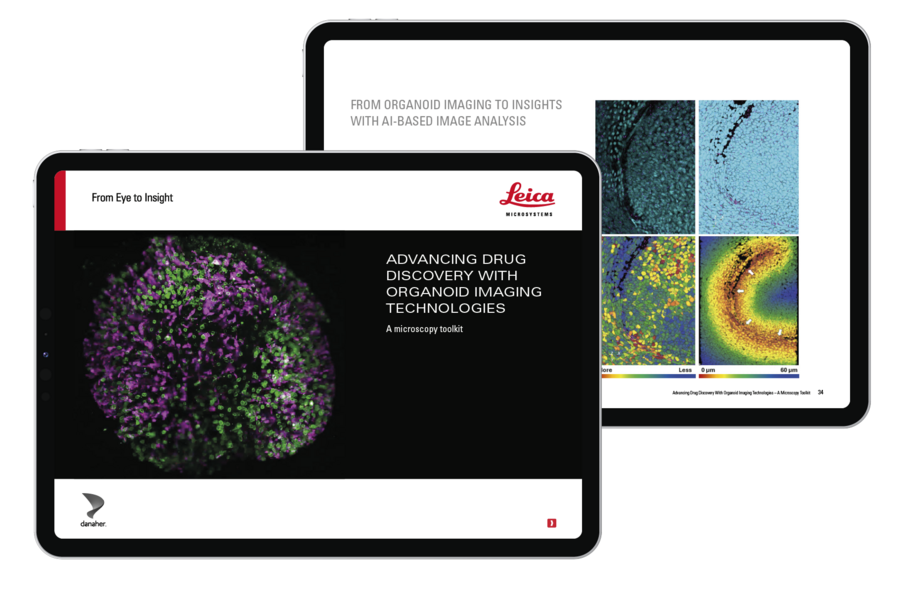
Why Download This eBook?
- Understand how to overcome key challenges when imaging dense 3D multicellular systems
- Read real-life case studies using microscopy to shed light on drug uptake, disease modeling, tracking single cell fate in organoids and spheroids, and more
- See how advanced imaging techniques can offer different insights into lung, intestinal, endometrial, and breast cancer organoids
- Discover tools to support every stage of your 3D imaging workflow – from monitoring organoid growth to AI-based analysis for more informed decision-making
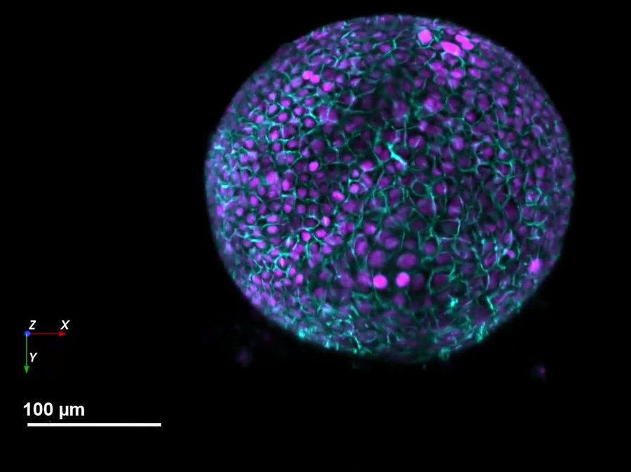
Murine liver organoid; nuclei - H2B-mCherry (magenta), cell membrane - mg-GFP (cyan). Courtesy of Franziska Moos, Liberali group, FMI, Basel, Switzerland.
Case studies include:
- Imaging lung organoids at the “liquid-air interface” to study potential therapeutic approaches for lung disease
- Investigating the emergence and maturation of secretory cells in intestinal organoids for spatiotemporal characterization of organoid development
- Advancing uterine regeneration therapies with endometrial organoids
- Imaging anti-cancer drug uptake in spheroids
The evolving landscape of drug discovery
3D culture systems such as organoids are seeing increasing use, driven by scientific innovation and regulatory shifts such as the FDA’s Modernization Act 2.0[1], which encourages the adoption of NAMs and NATs. The physiological relevance of organoids allows them to mimic human tissue architecture, offering insights into dynamic cellular interactions and spatial organization.
To fully harness the potential of these 3D models, specialized imaging approaches are needed to capture and quantify complex processes in real time. By enabling more predictive, ethical, and cost-effective research, organoids are helping to improve translational success in biomedical research, which will ultimately accelerate drug development.
Download eBook
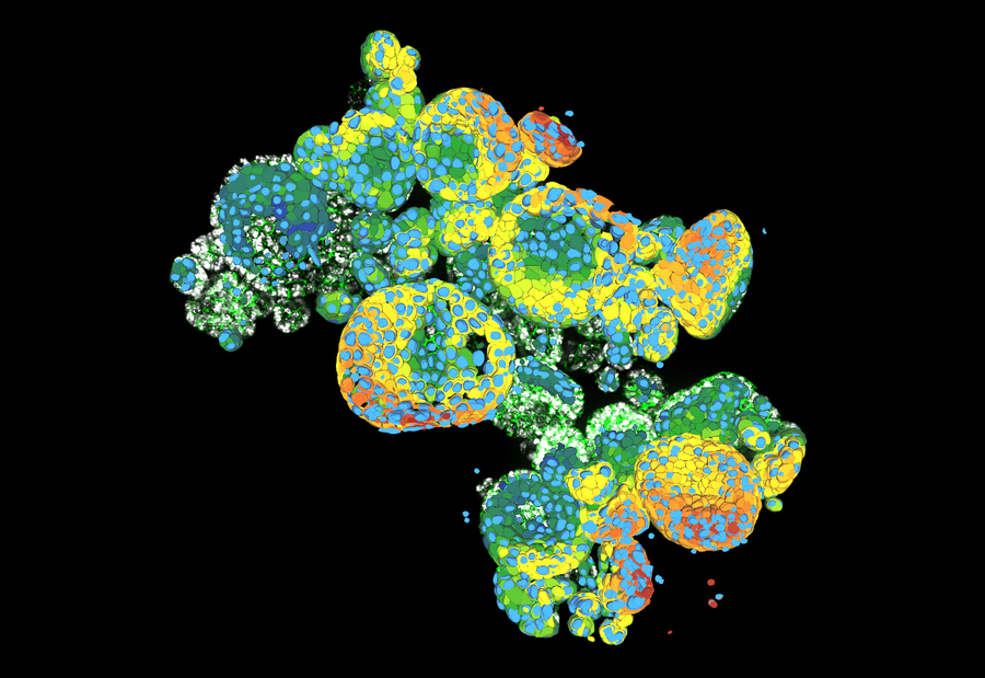
Organoid cluster labelled for nuclei (DAPI, blue) and plasma membrane (GFP, green). Thickness 100 µm. 469 Z planes were acquired using a THUNDER Imager 3D Cell Culture (63x objective) and then analyzed with Aivia. Courtesy M.Sc. Dana Krauß, Medical University of Vienna, Austria.
Organoids play an important role in our work, allowing us to model tumors in 3D and explore behaviors of engineered T cells in a way that’s both meaningful and patient-specific. Imaging is central to this as we aim to understand how cancer and immune cells communicate at their most intricate level, where every cell, structure, and interaction within a tumor can be visualized, understood, and ultimately targeted. We use advanced microscopy platforms from Leica Microsystems to explore cellular dynamics and spatial interactions
1. Zushin et al. (2023). FDA Modernization Act 2.0: transitioning beyond animal models with human cells, organoids, and AI/ML-based approaches. J. Clin. Invest, 133 (21); e175824. https://doi.org/10.1172/JCI175824.
