Live Cell Imaging Solutions
Our experts on live cell imaging applications are happy to help.
Your Live Cell Imaging Needs
To perform live-cell imaging experiments successfully, using the right approach is critical. When choosing a suitable microscope for your live‐cell imaging, the following 3 aspects should be considered: Specimen viability, image acquisition speed (temporal resolution) and required resolution in all three dimensions.
Specimen Viability
During live cell imaging, certain environmental conditions must be maintained to avoid detrimental physiological changes. In order to capture physiologically relevant cellular dynamics, live cell experiments require specific environmental conditions, including temperature, pH (via CO2), and humidity control. Furthermore, some experiments may even require hypoxic conditions. Modern incubation systems not only tightly control environmental conditions, they can also provide detailed data reports and alert users to temperature, gas, or humidity variations during the course of an imaging experiment.
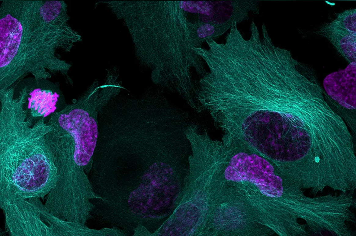
Light protection is another challenge for live-cell imaging because light exposure can influence living cells per se, as well as the fluorophore. For example, high light doses can lead to DNA damage or photobleach the fluorophores. To minimize or avoid the effects of photodamage, getting the right balance between sensitive detection, accurate label separation (if using >1 label) and the lowest light dosage for excitation is crucial.
Image Acquisition Speed (temporal resolution)
For live cell experiments, high speed acquisition is often critical, in particular for the study of fast dynamic processes such as vesicle observation. Using optical filters results in speed limitations due to the necessity for sequential imaging when changing filter sets for each color, used to study the interaction of multiple components. Gathering images sequentially requires more time than simultaneous image gathering and, as a result, rapid specimen motions can be missed during acquisition, as each color has a longer time interval from one image to the next. On top when the direct comparison between two or more colors is of essence, the signals may have moved even between the individual acquisition of the fluorophores, complicating the interpretation of the data.
Required resolution in all three dimensions
Multiple technologies are available for acquiring images in 3 dimensions over time. The choice of system depends on your experiment and whether higher speed or less sample illumination during imaging is your priority when acquiring the desired 3D resolution. Choosing the most appropriate system has traditionally required you to make a choice between a camera-based or confocal live cell imaging system, however modern solutions can provide both modalities in an integrated way.
Leica Microsystems offers the latest innovations in widefield and confocal imaging technologies for fast 3D live cell imaging and 3D live cell imaging over time (4D imaging) - with the THUNDER Imagers, Mica – the world’s first Microhub, and STELLARIS confocal platform.
These imaging solutions help optimize your study of live cells, even over long periods of time (e.g. days). They provide the necessary image contrast and resolution to facilitate the analysis of dynamic events. Based on the requirements of your live cell imaging experiments, you may need to prioritize high-speed acquisition, absolute correlation of labels without spatiotemporal mismatch or maximal 3D resolution. Leica provides a variety of live cell imaging systems to accommodate all of the above requirements and make sure that no key cellular events in your experiments are missed.
| THUNDER | Mica | STELLARIS | |
|---|---|---|---|
| Ease of use | + | +++ | ++ |
| Long-term live cell imaging | ++ | +++ | ++ |
| Fluorescence assays (wellplate) | ++ | +++ | ++ |
| Whole slide imaging (fluorescence) | +(+) | +++ | + |
| Fast live-cell imaging | +++ | ++ | ++ |
| Spatiotemporal resolution | +(+) | +++ | +++ |
| 3D Resolution | + | ++ | +++ |
| Upgradability/modularity | ++(+) | + | +++ |
+Basic, ++Standard, +++Premium
Request Demo, Quote, or Ask a Question
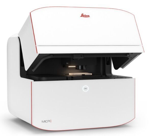
Mica.
The world's first Microhub
Unite widefield and confocal imaging in a sample protecting, incubating environment. With the simple push of a button, you have everything you need - all in one place - to supercharge your fluorescence microscopy workflows, power-up your research and streamline your path to results.
- Access for all
- No constraints
- Radically simplified workflows