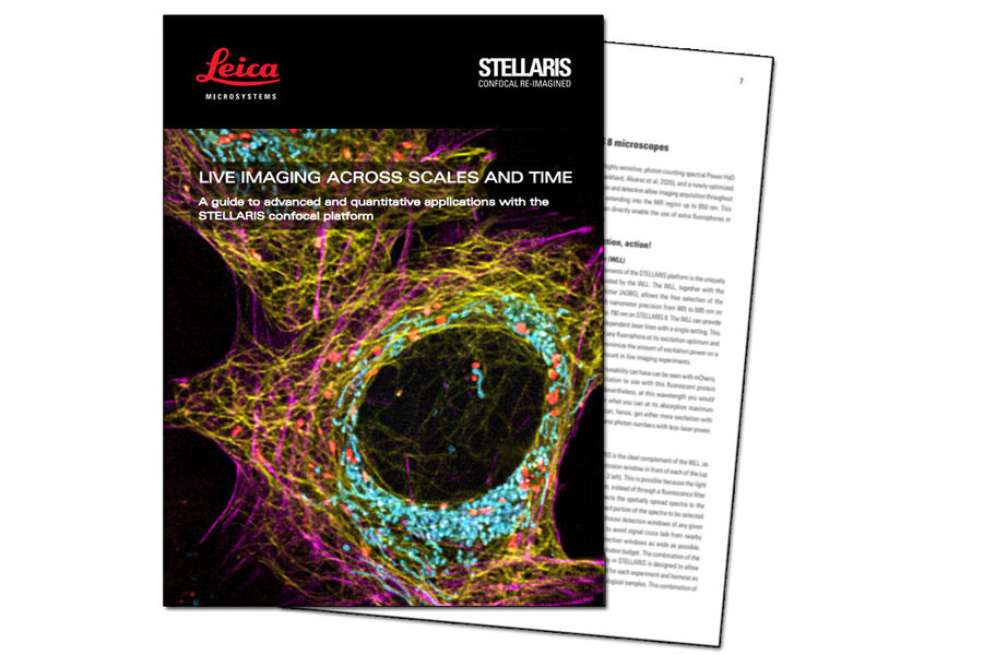
Live Imaging Across Scales and Time
Download the Guide for Advanced and Quantitative Life Science Applications
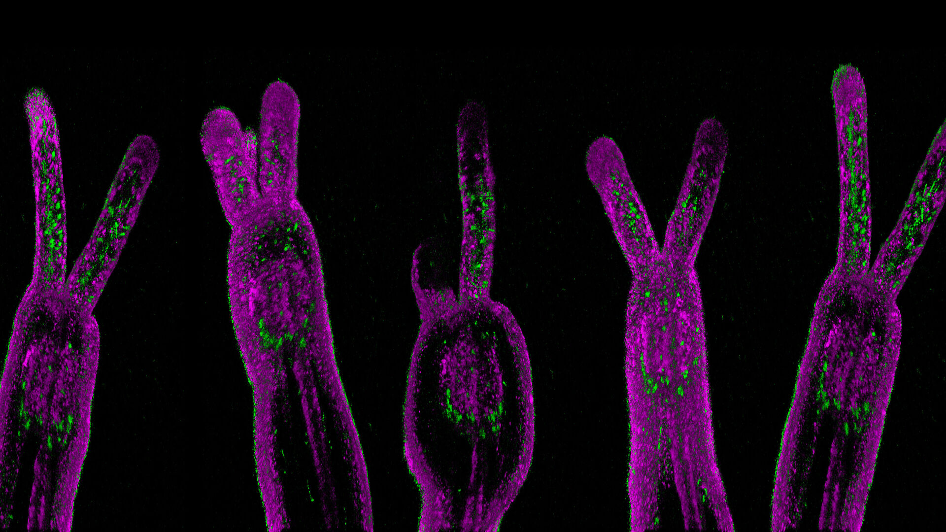
Take a journey through the biological scales. Go from proteins and molecules to cells and organisms. Explore from nanosecond signals to processes over hours or even days and from spatial distributions to readouts linked to the microenvironmental changes in cells.
For all of these mechanisms, the STELLARIS confocal microscope provides ways to help you investigate and generate insights.
Get the Live Imaging Guide and find out what is possible in live imaging with a STELLARIS confocal microscope.
STELLARIS advanced imaging methods for your research
Confocal microscopy | Multiphoton microscopy | Lightsheet microscopy | STED microscopy / Nanoscopy | Lifetime-based imaging
STELLARIS live imaging is suitable for
Cell biology | Cancer research | Virology | Immunology | Developmental biology | Neuroscience | Plant biology and many more...
Get the Live Imaging Guide and find out what is possible in live imaging with STELLARIS.
