为活细胞成像创造新选择
使用THUNDER与Aivia
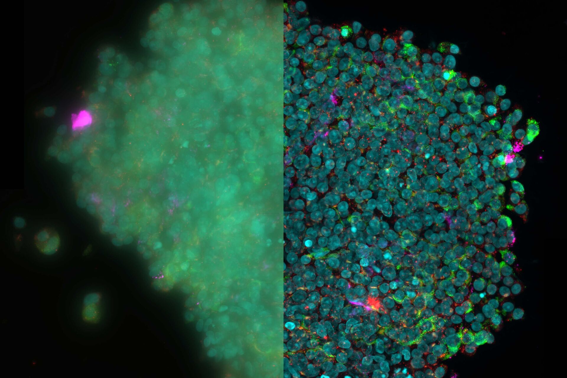
![[Translate to chinese:] 3D reconstruction of an isolated human islet THUNDER-Imager-3D-Cell-Culture_isolated-human-islet.jpg](/fileadmin/_processed_/d/1/csm_THUNDER-Imager-3D-Cell-Culture_isolated-human-islet_9d5da59242.jpg "[Translate to chinese:] 3D reconstruction of an isolated human islet")
对厚实的活体样本进行成像时,主要挑战之一是获得图像质量与组织完整性之间的平衡。长时间的图像采集期间,弱信号光会导致低信号水平,导致图像对比度低以及分割和分析困难。需要通过高剂量成像或高时间分辨率成像技术加强信号强度时,这一问题更加突出。一个常见问题是:我如果快速成像、一次完成,会不会造成样本过度漂白或者细胞死亡?
新人工智能系统可以有效解决这一问题。THUNDER和Aivia这两种新技术可以在不影响样本存活时间的前提下,获得高质量的图像和宽场图像分析结果。THUNDER使用计算机方法显著提高弱光成像时的图像对比度。Aivia基于机器学习的目标检测可以分割并精确分析这些更高对比度的图像,生成高度复杂样本的可靠重建结果。
相关文章
-

A Meta-cancer Analysis of the Tumor Spatial Microenvironment
Learn how clustering analysis of Cell DIVE datasets in Aivia can be used to understand…
Apr 26, 2024Read article -
 tissue.")
Mapping the Landscape of Colorectal Adenocarcinoma with Imaging and AI
Discover deep insights in colon adenocarcinoma and other immuno-oncology realms through the potent…
Apr 26, 2024Read article -

Spatial Architecture of Tumor and Immune Cells in Tumor Tissues
Dig deep into the spatial biology of cancer progression and mouse immune-oncology in this poster,…
Apr 26, 2024Read article
相关页面
-
-
THUNDER Imaging Systems
为了解答重要的科研问题,这些系统甚至能深入原始样品中实时呈现清晰的细节,不会产生任何离焦模糊。现如今,为3D样品进行清晰成像就像使用您最喜爱的摄像头荧光显微镜一样简单。采用 Computational…
Visit related page -
想了解更多信息?
请咨询我们的专家。
您想获取专人咨询吗? Show local contacts