
Challenging immunosuppressive resistance
Immune checkpoint blockade (ICB) therapies have demonstrated great clinical benefit in a variety of malignancies, but there are many patients that do not respond. Despite many preclinical and clinical studies, the mechanism of ICB is not completely understood. Development of novel therapies targeting other immunosuppressive mechanisms or combining ICB with conventional chemo/radio therapies has the potential to increase the number of patients that achieve durable responses. However, the optimal therapy or combination of therapies for a patient is likely influenced by which immunosuppressive mechanisms exist within the tumor microenvironment (TME) and the phenotype and function of tumor-infiltrating immune cells. To delineate the mechanisms of immunosuppression we selected FFPE tissue sections from three syngeneic murine cancer models and performed tumor-immune profiling to spatially understand the contributions of both immune cells and cancer cells. Mouse reactive antibodies targeting specific cellular populations will help identify the location of immune cells within the tumor site and can be used for proof-of-concept studies. This approach can lead to novel insights to synergize anti-tumor activity of ICB therapies and will help to identify differences between responders and non-responders.
Unlocking tissue insights with open multiplexing solution
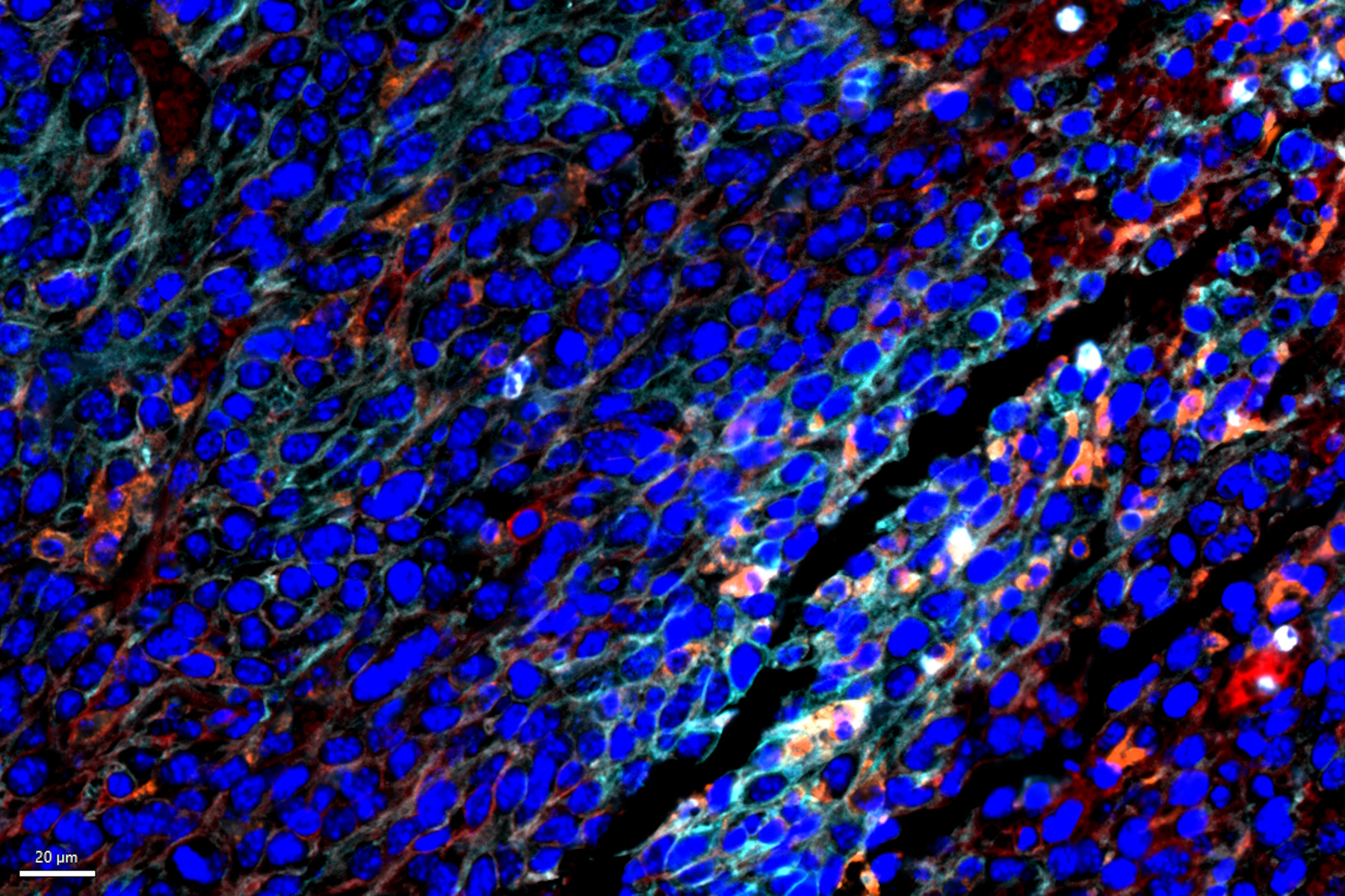
Cell DIVE Multiplex Imaging Solution allows probing and imaging of dozens of biomarkers on a whole single tissue section with an iterative staining and dye inactivation workflow. At its core, Cell DIVE is a precise and adaptable open multiplexing solution that enables flexibility in antibody selection of biomarker panels used in a multiplexed imaging study. Cell Signaling Technology (CST) has a broad portfolio of IHC-validated antibodies to detect key proteins in the TME, enabling immune cell detection and phenotyping in tissue. CST offers off the shelf (OTS), ready-to-ship antibody conjugates that have been verified to work on Cell DIVE and offers custom conjugation of IHC-validated antibodies to fluorophores and other detection reagents. CST employs a rigorous approach to IHC validation, followed by verification on the Cell DIVE platform to ensure successful detection of proteins. Here, we demonstrate multiplexed Cell DIVE imaging using a novel panel of dozens of CST biomarkers across multiple mouse syngeneic tumor types. In summary, multiplexed whole slide imaging using validated antibodies and Aivia AI-based image analysis software enables spatially resolved tissue analysis of the tumor microenvironment, including new insights into spatial biology.
")