Anatomic Pathology Microscope Solutions
Working in the highly specialized field of anatomic pathology requires not only a wide knowledge and understanding of the pathological and clinical aspects of many diseases; it is also essential to work efficiently and confidently under pressure.
Expert anatomic pathologists need to be able to rely on high-quality microscope imaging that shows clear differentiation of tissue structures and cellular details for accurate results. And with many hours spent each day working at a microscope, achieving ergonomic comfort is an additional priority to help reduce physical strain and maintain mental focus.
Contact us to learn how Leica pathology microscopes can support efficient and accurate diagnoses.

What is anatomic pathology?
Anatomic pathology is the medical study of tissue samples obtained from surgical biopsies or autopsies. Anatomic pathologists investigate structural changes within organs or tissues, both as a whole and microscopically within the cells.

What are microscopes used for in anatomic pathology?
Microscopes help in the examination of stained tissue specimens or cells and can reveal abnormalities. Histological or dermatological specimens typically stem from surgeries or biopsies, are frozen or fixed in formalin, sliced thinly, and cover-slipped in preparation for microscopic analysis. In a sister discipline to surgical pathology, cytopathologists look at cells which have been taken from body fluids or acquired through scraping or aspiration. Dermatopathologists are specialized in the examination of skin.

What disciplines are covered under the umbrella of anatomic pathology?
Anatomic pathology encompasses various disciplines including histopathology, cytopathology, dermatopathology, and forensic pathology. Together, these branches work towards building a comprehensive and ever-increasing picture of anatomic tissue and cellular structures to improve patient health.
Our Solutions for Anatomic Pathology
DM1000 LED | Visoria B | DM3000 & DM3000 LED | |
| Occasional use | x | - | - |
| Average to high workload | - | x | x |
| Adjustable both microscope height and viewing height & angle with eyepieces | x | x | x |
| Control knobs in close proximity with symmetrical layout | - | x | x |
| Adjustable height of stage and focus controls | x | x | x |
| Adjustable torque for focus knobs | - | x | x |
| Multiple viewing system for simultaneous observation by several users | - | x | - |
| Fast objective change with toggle mode | - | - | x |
| Automated illumination manager | - | x | x |
| Robust compact design | x | x | x |
| Go to product page | Go to product page | Go to product page |
x = included, - = not available
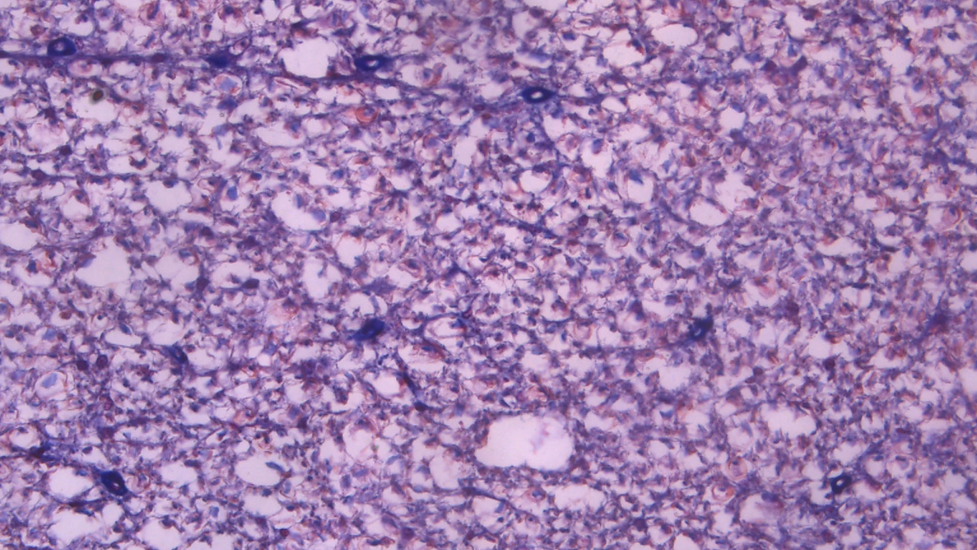
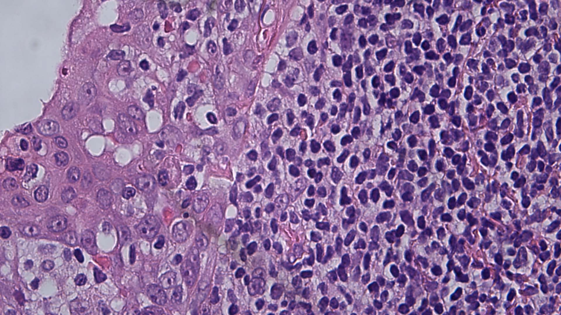
Speed Matters in Histopathology
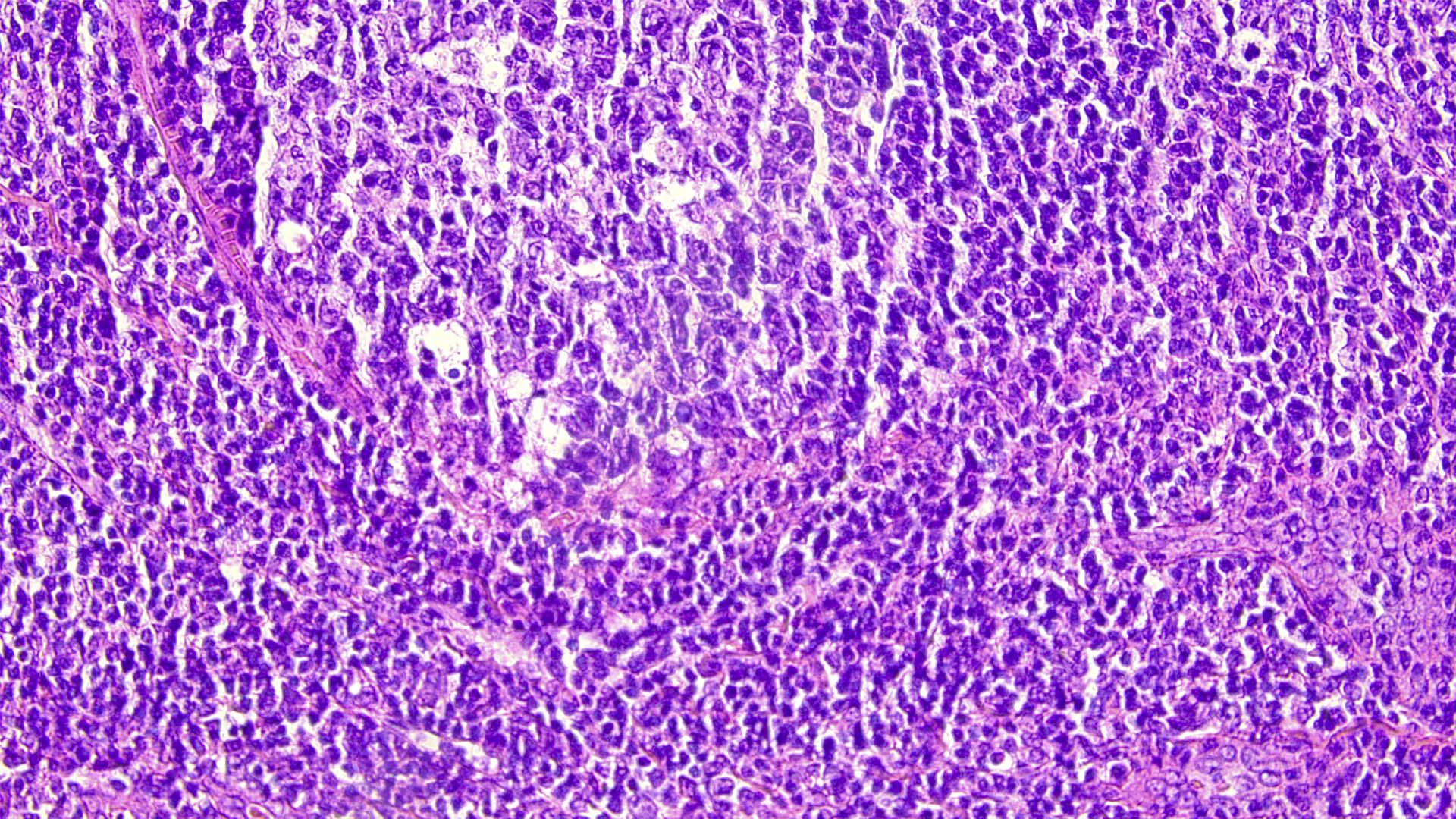
In the discipline of histopathology, also known as surgical pathology, time is of the essence to spot inflammatory diseases, abnormal growths, infections and cancer in surgical tissue samples and biopsies. There are cases where the tissues need to be examined while the patient is still under anesthetic in the OR, all in a matter of minutes.
As the pathology results will impact what happens next on the operation table, a high level of concentration is needed to look for the presence of disease, with a comfortable workstation and an efficient workflow to reduce physical strain. Microscope images also need to be crisp and clear, so that pathologists can confidently make their diagnoses.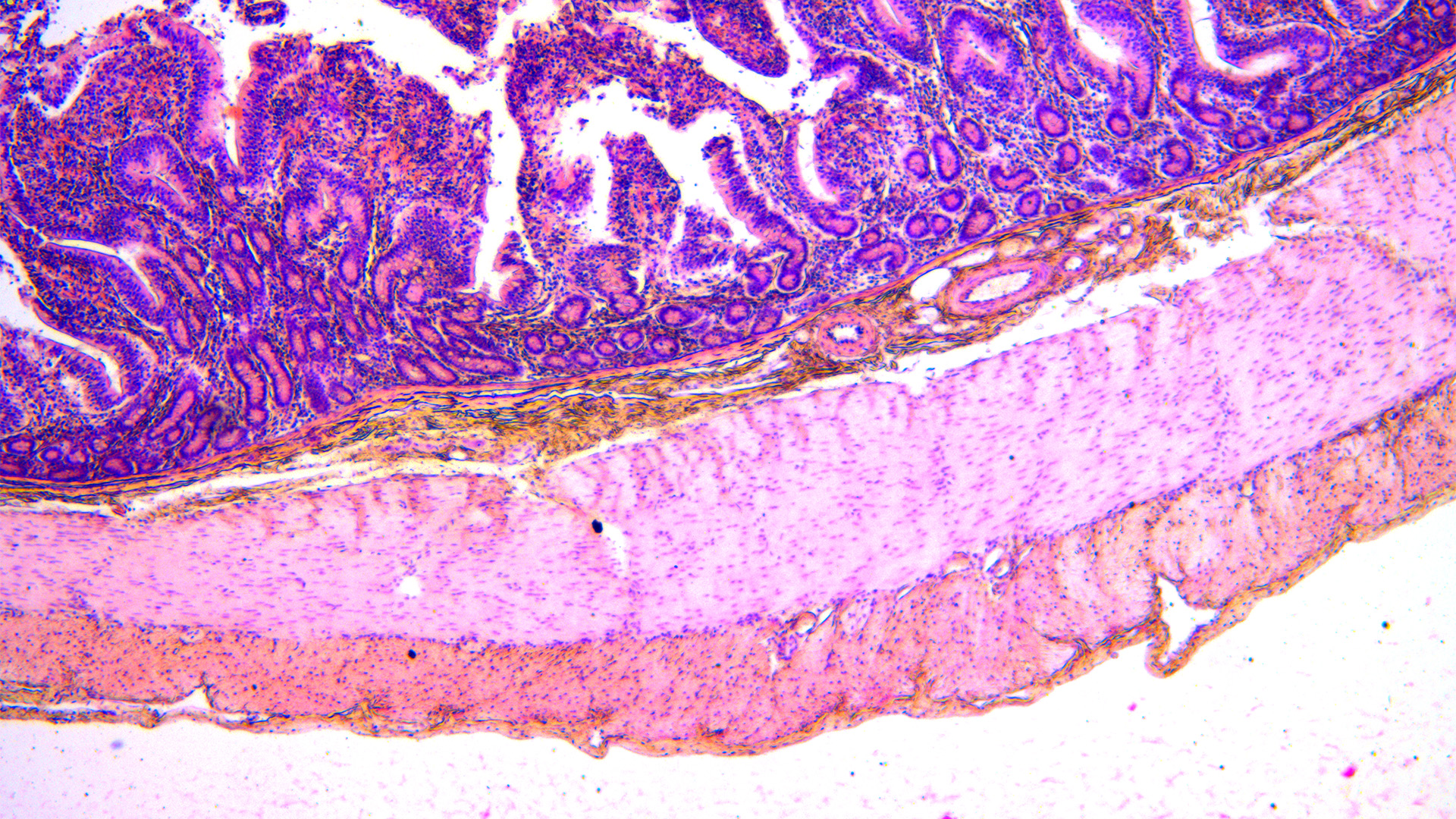
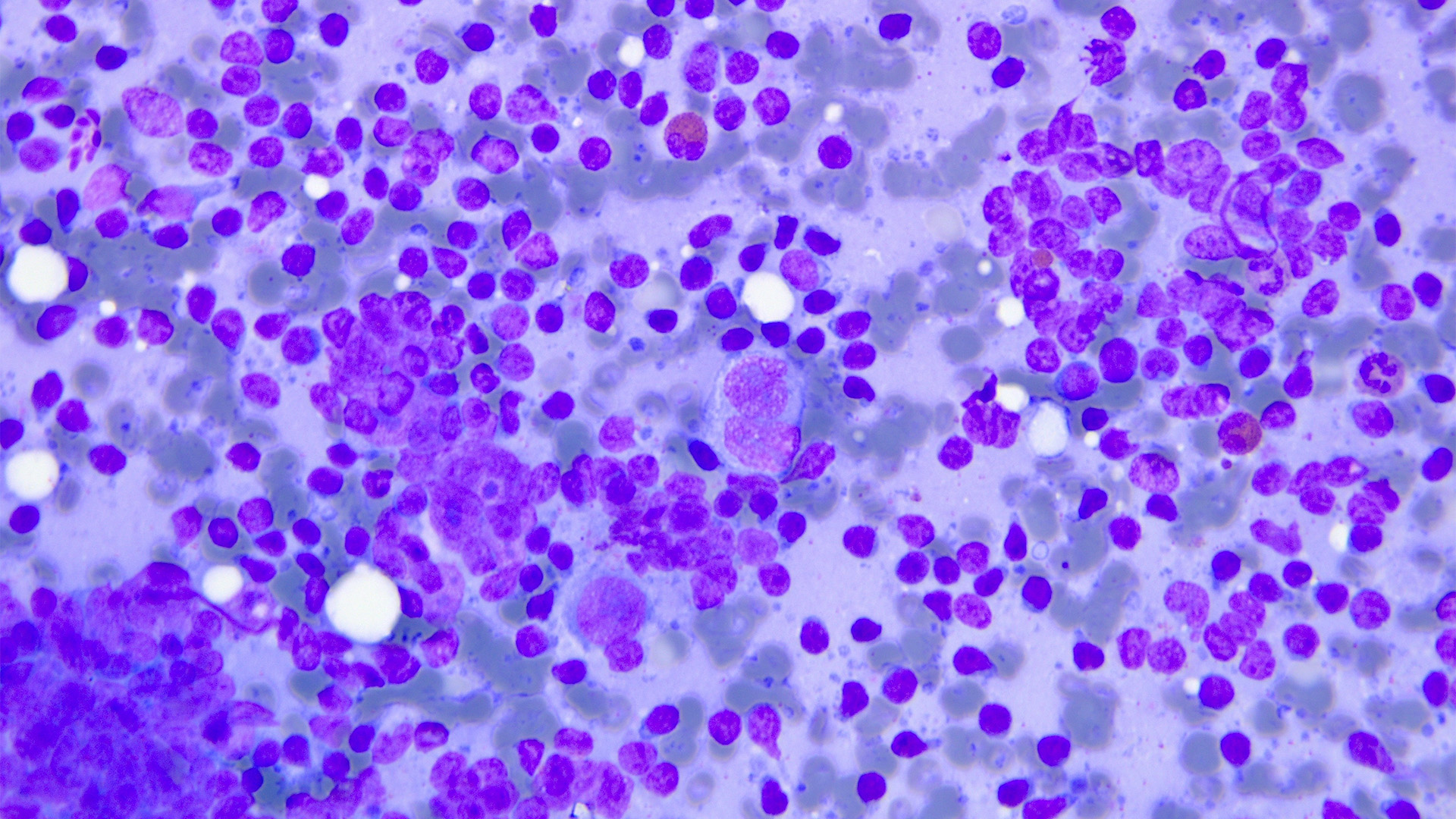
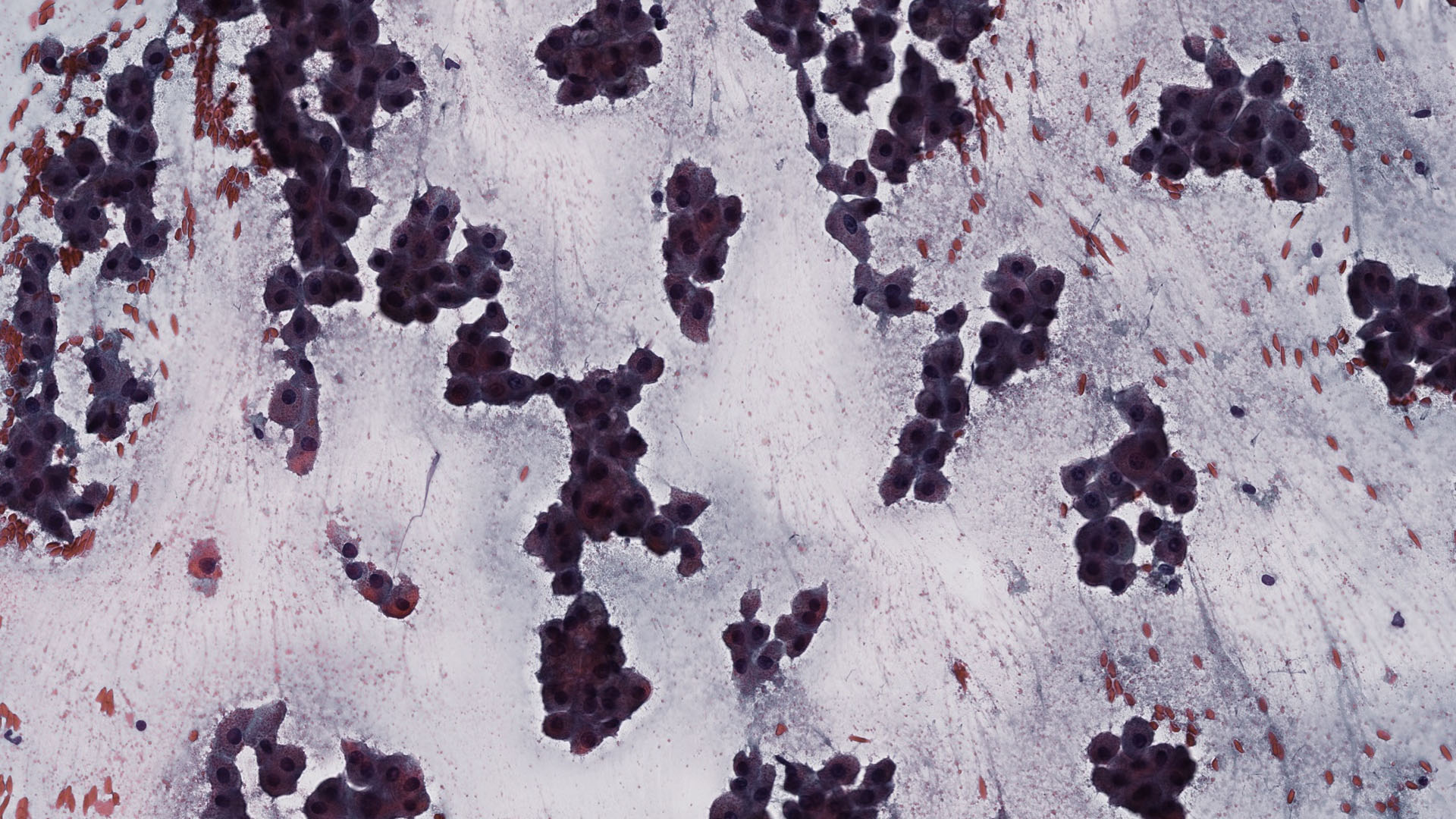
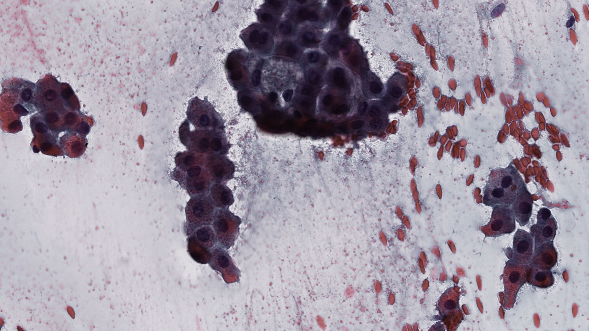
A Closer Look at Cytopathology
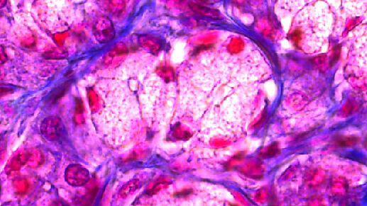
Unlike histopathology, which involves examining an entire section of tissue, typically containing many types of cells, cytopathology generally involves looking at individual cells or clusters of cells taken from a tissue sample.
Microscopy plays a pivotal role in this work by enabling the clear visualization of cellular structures and abnormalities. Through microscopic examination, e.g., pap smear tests, cytopathologists can identify cancerous cells, assess their morphology, and determine their behavior, aiding in accurate diagnosis, prognosis, and treatment planning for patients.
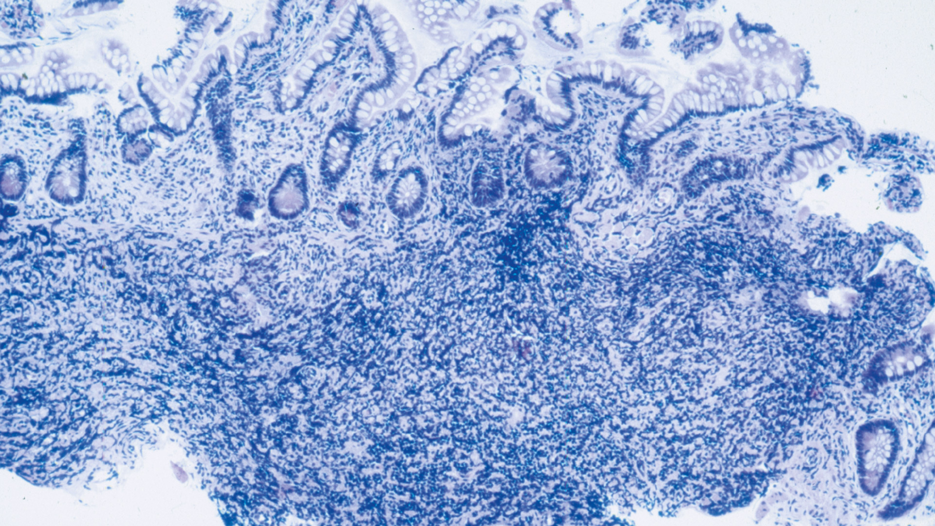
Diagnose With Confidence
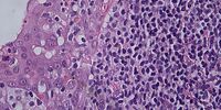
Tissue sections are typically stained with Hematoxylin and Eosin (H&E). Different parts of the specimens take up varying degrees of color, so that the different color shades and intensities provide insight on abnormalities.
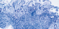
If specimens are not stained and for example observed with phase contrast, the difference in contrast will give pathologists insights about variations in structure.
The optical quality of the objective is a main factor that influences how well pathologists can discern colors and structures in their specimens.
Great Color Fidelity
Leica pathology microscopes are available with three groups of objective classes differing in quality of chromatic correction. When used with color and field-flatness corrected objectives, the image you see shows the specimen as it is, untainted from optical aberrations, such as color fringes or distortions. Thus, you can make diagnoses confidently on the basis of what you see.
Cameras suitable for the Leica pathology microscopes capture what you see in true colors and provide high-quality images for your reports. The images can be archived in the hospital information system thanks to the TWAIN capability of the cameras.
Minimize the Strain
High workload, time pressure, long hours on the microscope – all this may strain pathologists’ muscles, making them feel tense. With time, strain and tension may take their toll, impacting their work and personal life.
Therefore, pathologists should take any physical discomfort seriously and be concerned about muscle tension resulting from their work posture or repetitive movements.
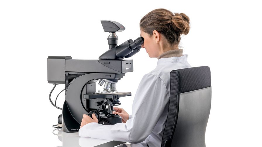
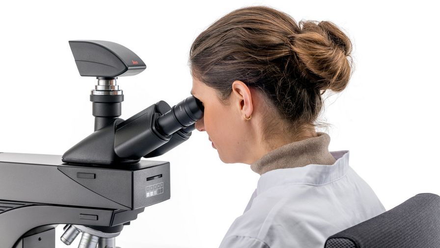
Work in Comfort
Ergonomic design in microscopes helps prevent strain in the back, neck, arms, and hands.
You can adjust your Leica pathology microscope to suit your body height and preference as a right- or left-hander. You can also choose the viewing angle of the oculars as well as set the focus knobs to the optimal position for your hands.
This helps you to achieve a more relaxed working posture, with aligned shoulders and symmetrical operation of the stage drive and focus.
In addition to that, ergonomic features can have a direct impact on efficiency: automatic switching between two objectives, the so-called toggle mode, reduces repetitive movements and contributes to efficient work.
Frequently Asked Questions Anatomic Pathology
Yes, it does. As soon as you use an ergonomic microscope, you will feel the difference immediately, not only in your posture, but also in the workflow. Leica clinical microscopes help you save time with a design that minimizes unnecessary and repetitive movements that can cause strain and fatigue.
Accessories, such as the Enersight software platform for fast and easy image acquisition and sharing and selected cameras with their TWAIN capability to archive images in the hospital or lab information system, help integrate the microscopes more smoothly into your workflow.
Yes, Leica Microsystems offers a variety of ergonomically designed tubes, so that users of different body size can sit upright in a relaxed posture. Users can operate the stage with their left or right hand and adjust the height of the focus knobs to suit their hand size and arm position. Further ergonomic accessories, such as ErgoTubes, ErgoModules, and ErgoLift, are available.


