Software Tutorial Videos
You want to learn more about the LAS X, Enersight, Aivia or need advice on its application?
Visit our YouTube Channel!
There you will find numerous videos with helpful tutorials as well as tips and tricks on how to use our microscope software.
An all-encompassing solution
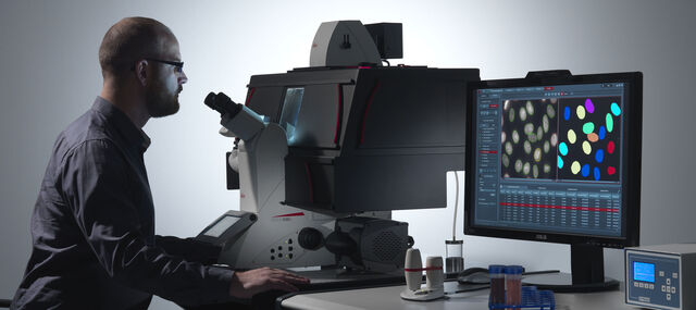
With an intuitive user interface and straightforward navigation, our microscope imaging software guides the user through their workflow, whether fast image acquisition or sophisticated expert analysis. A range of specific modules allows configuration of the microscope as a dedicated high-performance tool for almost any application.
LAS X encompasses all microscope solutions for Life Science and Industry applications, offering maximum flexibility. The previous Leica Application Suite continues to be supported.
Core Features
- Microscope and digital camera configuration and control in a fully integrated manner.
- Basic annotation tools allow image and calibration data to be added to images.
- Auto and manual exposure adjustments allow optimized imaging conditions.
- A thumbnail gallery of acquired images, which can be reviewed quickly and easily.
- Automatically calibrated images using data read from Leica microscopes and cameras; a scale bar indicates image size.
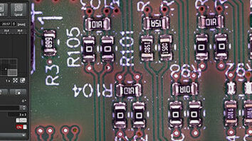
LAS X Industry
- Observe your sample over the full screen
- Make streamlined and flexible measurements when doing analysis
- Enhance your imaging with stitching in X, Y, and Z
- Acquire and recall reliable and reproducible results
- Customize report templates for your needs
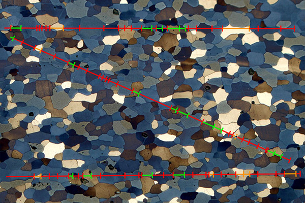
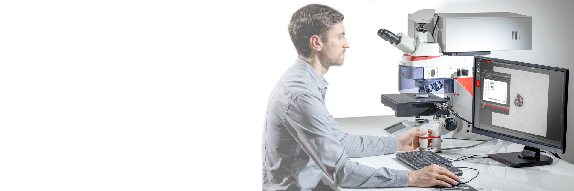
LAS X Materials Science Modules
- Allows users to rapidly perform structural analysis of steel and other materials.
- Enables the advanced analysis of multiple phases and microstructure components.
- Specifically developed to combine accurate object and area measurements with high flexibility to tailor automated analyses to a user’s particular needs.
- Offers a significant improvement in ergonomics and aids users in maintaining consistent accuracy in measurements over time.
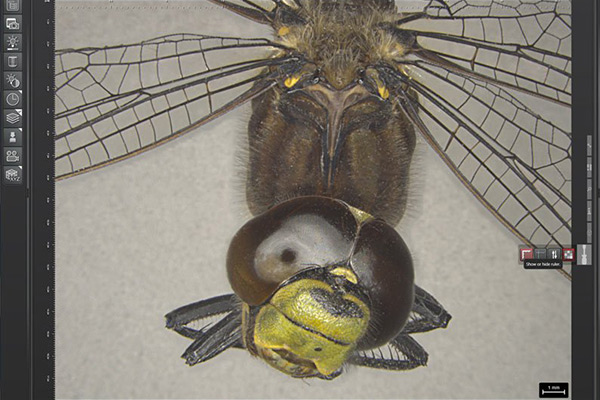
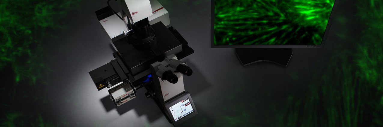
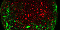
LAS X Life Science
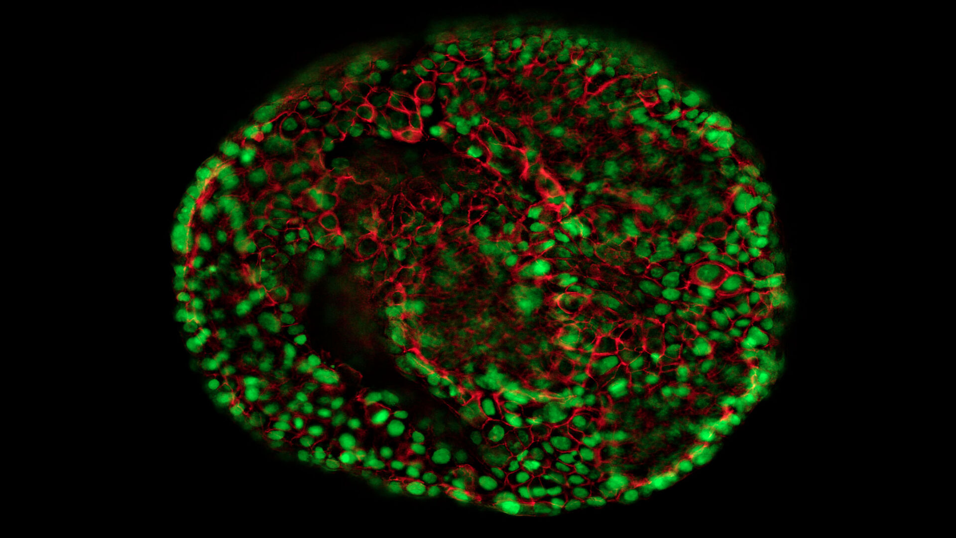
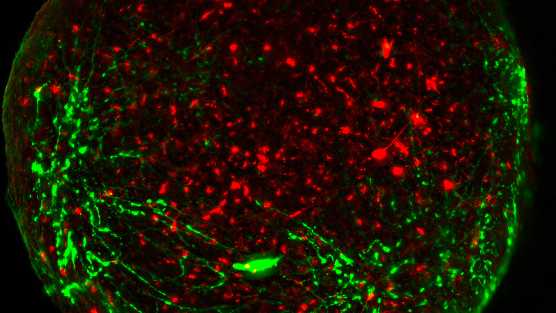
- Integrates confocal, widefield, stereo, super-resolution, and light-sheet instruments from Leica Microsystems.
- Create fast overviews and identify the important details instantly with the LAS X Navigator - a GPS for your cells
- Imaging tasks become intuitive allowing to focus on your research
Frequently Asked Questions Microscope Software
The Enersight software can be operated directly on a monitor, mobile device, or with a computer. You can use Enersight On-Screen Display, which is embedded in the hardware of all compatible devices, to operate your microscope or camera after plugging in the power supply, USB stick, mouse, and monitor. To use Enersight Desktop, a PC-based software, download it from the Leica website and connect the device via a USB-C/A cable to a computer. Alternatively, you can use your mobile device to control the microscope or camera via the Enersight Mobile App. To connect a mobile device to a camera, please refer to tutorial clip First steps with Enersight.
The Ivesta 3 (Integrated camera) microscope, Flexacam i5 and c5 microscope cameras, Flexacam C3 camera, and Emspira 3 digital microscope work with Enersight.
With Enersight software platform, you can acquire images, record videos, compare a live image with a reference via one click, perform measurements, annotate live images, create, and use custom overlays, share images, and benefit from user management functionalities that allow you to tailor the Enersight User Interface to your needs.
Life image and video recording in 4k, 1080, or 720 resolutions with up to 60 fps (frames per second) in MP4 format. Image acquisition in 4k, 1080, 720, 12MP or 3MP resolutions with a JPG/JPEG, TIFF, BMP formats.
First, navigate to the Measurement Tab and Calibration, then place a scale on your microscope stage and set the zoom to the lowest value (e.g., 0.6x), click on Create calibration, enter a value for the linear bar in the desired units, and click "Ok". The calibration value for this magnification is now saved. Then, move to the next zoom value (e.g., 1.0x) and repeat the same actions. You can do the calibration for multiple, or all zoom values if needed. A list of all your saved calibration data can be found in the measurement tab. While doing measurements, please make sure your zoom value corresponds to the calibration value you saved for this magnification step.
To create a custom overlay, please go to the Measurement tab, draw the desired shapes, or perform measurements, then save it as an overlay by clicking on the corresponding button. Your overlay will appear in the Overlay tab under Custom overlays. To use it during live imaging, simply click on it. To upload overlays from the computer, click the Upload button in the Overlay Tab. To edit overlays, navigate to the Measurement tab and click on Import annotations. The window with custom overlays will pop up, then select the desired overlay which will be shown in the measurement tab in an editable format. To save your changes, save this overlay again. To use custom overlays for multiple devices and operation modes, simply save it onto a USB stick.
Using Enersight On-Screen Display, you can set up a network folder in which you can save your images. Please refer to tutorial clip Documentation and Sharing. To be able to save files on a network folder via Enersight Desktop, please set the network folder as default in the Gallery tab. To be able to upload files to a network folder via Enersight Mobile, please use your mobile device’s OS capabilities, by clicking on the Share button in the triple-dot menu of your image or video file.
Yes, you turn the live image into a negative using the camera settings. Take a picture, set it as a comparison in the Gallery, change the transparency to the preferred level, and click on Comparison in the menu bar. As a result, two images will appear on the screen. To overlap one image with another, double click twice on the screen. The comparison functionality is available with Enersight OSD and Enersight Desktop.
In the Measurement tab of Enersight, you have the following tools: selection, distance line tools, measurement tolerancing*, angle, circle tool, edge detection (snap-to-edge)*, shape / polygon, point-to-point measurement, consecutive numbering, and text / line / arrow annotations.
*only available with Enersight Desktop and Enersight OSD
Open Enersight OSD and go to the “Network” tab in the settings. Connect your camera to the network in the following ways:
- via LAN cable with DHCP turned on or manually setting the IP address provided by your IT support; or
- insert a Wi-Fi dongle into the back, use client mode, scan the SSIDs of your Wi-Fi, select the one you want, enter your username and password and turn on the Wi-Fi.
For information on how to set up the network folder or send pictures by e-mail, see the Youtube video tutorial “Documentation & Sharing”.
The only system requirements for Enersight Desktop are using Windows 10 Pro or Windows 11.
The licenses can be checked up in via the CodeMeter control center (included in the LAS X software package). After having opened the control center, click on WebAdmin. There you will find your licenses. In case you use a hardware dongle, it has to be inserted into the PC.
In case you have a licensing request or you would like to extend the validity of a demo dongle, we require the context file of your dongle. This can be generated by the CodeMeter control center (including the LAS X software package) and contains all relevant information regarding the dongle and the corresponding license modules. Download the instructions.
The context file can be generated by the CodeMeter control center (including the LAS X software package) and contains all relevant information regarding the dongle and the corresponding license modules. Download the instructions.
A dongle is the location which contains the licensing file and therefore licenses the software. This licensing can either be located on your PC (soft dongle) or on a special codemeter USB stick (hard dongle). The hard dongle offers a Plug&Play function and simply has to be inserted into the USB port of the PC which should be licensed.
LAS X software only runs with Windows, but for a MAC we have a dedicated software called Leica Acquire that you could download for free from the Apple store:
https://apps.apple.com/it/app/leica-acquire/id733706983?mt=12
There is no software for Linux.
- Find your LAS license number as well as the order number on the software box that is enclosed to the system.
- Find your LAS license number as well as the order number on the microscope system on a licensing label, in case the instrument was bought after 2017-04-01.
- Find your order number on your invoice.
- Your license number (LASxxxxx) is printed on a label attached to the LAS DVD packaging.
Find a detailed description of LAS X and its modules on the product page.
Please start the online activation again. For this purpose, you require your LAS license number and your order number.
Please start the online activation again. For this purpose, you require your LAS license number and your order number.
Start the online registration again with the assistance of the online registration tool, which is included in your software package. For this procedure, your require your LAS license number and your order number.
You will find your PC Site Code when you click the 'Start' button in Windows, "All programs" > "Leica Applications Suite" > "LAS License for Activation and Registration".
Afterwards, choose the button "Start LAS for license Activation and Registration".
Please start this program. Within the program, select the button "Options" > "Registration and Activation".
Here, you can see the Site Code now.
The PC Site Code is a clearly specified identification number of your PC that will be defined by the implemented hardware.
You will find your LAS license number as well as the order number on the software box that is enclosed to the system.
Furthermore, you will find your LAS license number as well as the order number on the microscope system on a licensing label, in case the instrument was bought after 2017-04-01.
You will find your order number on your invoice.
Download manual "how to activate an offline-PC."
Download manual "how to activate an online-PC".