
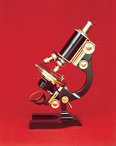
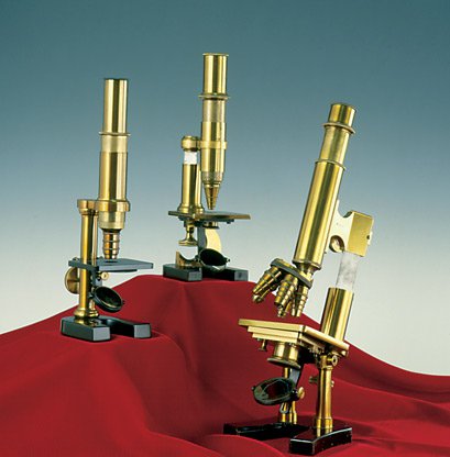
A History of Success
A historically close collaboration with the scientific, medical, and industrial communities is the key to Leica Microsystems’ tradition of innovation. Drawing on users’ ideas and developing solutions tailored to meet their requirements, the company continually sets new standards in markets it serves. This also applies to the services Leica Microsystems provides customers with: The company offers expert technical services, but also application support for all application-related questions.
2025

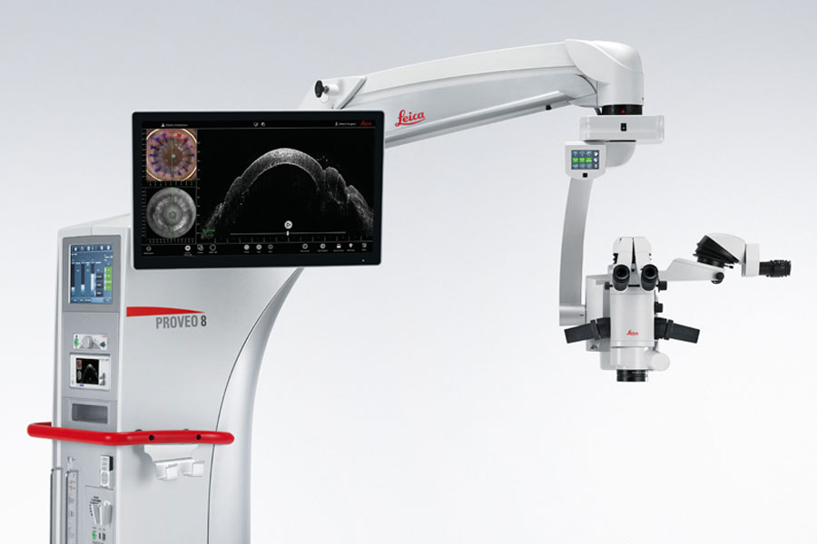
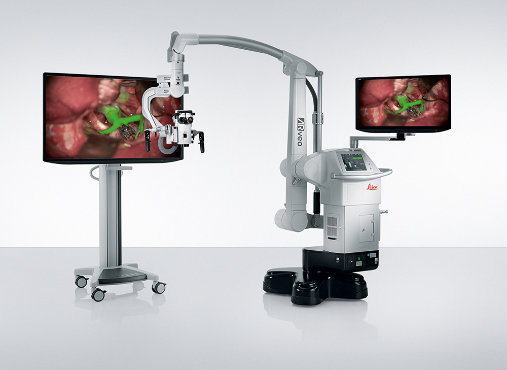
Proveo 8x shapes the future of ophthalmic surgery with real-time 3D visualization
Ophthalmic surgeons are empowered with the Proveo 8x digital microscope to operate with unmatched comfort . Well-informed decisions and efficient workflows are enabled by real-time 3D imaging , flexible visualization setups , and seamless data integration from Phaco and OCT . Surgeons are supported by an immersive view with accurate colors and texture representation , as well as depth resolution , regardless of the viewing options used.
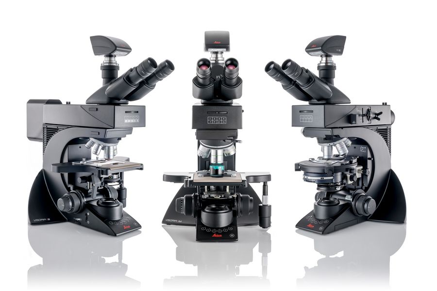
Visoria upright microscopes enhance comfort and efficiency in routine microscopy
With the launch of the Visoria series, Leica Microsystems enables users in clinical, industrial, and life science settings to perform routine microscopy tasks with more comfort and efficiency. Thanks to their ergonomic design, encoded functions, and digital viewing options, the Visoria upright microscopes help reduce strain and save time for users when examining histological samples, inspecting materials, or analyzing geological structures.
Leica Microsystems expands imaging workflow capabilities with the acquisition of ATTO-TEC
By integrating ATTO-TEC fluorescent dyes and reagents, Leica Microsystems now supports researchers throughout the entire microscopy imaging workflow, from sample preparation to advanced analysis . This allows for more reliable and reproducible results in a variety of scientific fields. One notable example is high-plex 3D imaging in cancer research, where this combined expertise helps scientists accelerate therapy development by revealing the invisible.
2024

Celebrating 175 Years of Leica Microsystems
The history of Leica Microsystems is closely related to the world-renowned brands Leitz and Leica. It is defined by innovations that reveal the invisible and empower our customers to create a better and healthier world. The origins of the company date back to 1849 when a family business for optics was founded in Wetzlar, Germany. For 175 years, Leica Microsystems has helped to shape the future and continues to do so today with the latest digital innovations that provide new insights for research, medicine and industry.
UC Enuity is the next-generation ultramicrotome that saves valuable time and resources through its automated setup functions, providing state-of-the-art sectioning quality
UC Enuity lets users experience the future of ultramicrotomy with autoalignment of block face and knife, and auto trimming. Using fluorescence or µCT data, UC Enuity can trim a target to deliver high-quality sections for volume electron microscopy experiments.

The evolved ARveo 8 Digital Visualization Microscope for Neurosurgery is the future of digital surgery
With the evolved ARveo 8 surgeons can experience a new level of neurosurgical visualization. Its clinical 3D application transforms brain tumor surgery visualization. Together with the all-in-one surgical visualization headset, MyVeo, users benefit from a single, integrated view of clinical data right in front of the eyes.
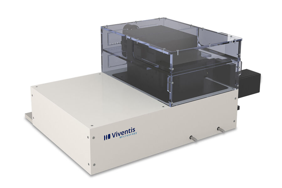
With the integration of Viventis Microscopy, Leica Microsystems adds cutting-edge light sheet solutions to its portfolio
The Viventis LS2 Live light sheet fluorescence microscope combines multi-view and multi-position light-sheet imaging to illuminate life in its entirety. Scientists can benefit from deep, long-term imaging that reveals the intricate details and dynamics of biological systems.
2023
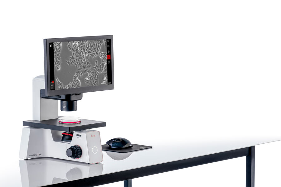
Mateo TL helps to improve the reproducibility of routine cell culture checks
For researchers who require consistent experimental outcomes, the Mateo TL digital transmitted light microscope enables consistent measurement of confluency, increasing confidence in the success of downstream experiments. User-friendly and intuitive, Mateo TL assists all lab members to check and document cell growth status through objective confluency evaluation across individuals or experiments.
2022
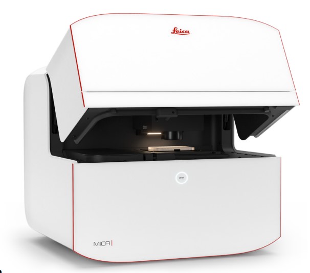
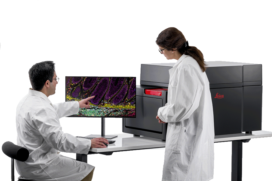
Mica, the world's first Microhub, makes spatial context accessible for all life science researchers
Mica is a type of wholly integrated imaging solution that leverages machine learning software, automation tools and unique fluorescence unmixing techniques to automate the imaging workflow for researchers, regardless of their microscopy experience levels. Mica offers users the ability to visualize four labels simultaneously in both widefield and confocal using Leica’s patented FluoSync technology, which offers four times more data with 100% correlation compared to traditional fluorescence imaging methods.
Coral Cryo workflow solution with high precision confocal 3D targeting
The Coral workflow solution, which includes a STELLARIS 5 Cryo confocal microscope with cryo stage and shuttle, enables researchers to increase their success rate in cryo-electron tomography experiments. The Coral Cryo workflow solution contains three steps: plunge freezing, cryo light microscopy and targeting with the CryoET software. The position of the area of interest is then exported to the electron microscope (EM) for precision targeting at the next step.
2021
Aivia drives research with autonomous image analysis in microscopy
Powerful artificial intelligence (AI)-guided image analysis combines with visualization solutions for data-driven scientific discovery. Aivia is an innovative and complete 2-to-5D image visualization, analysis, and interpretation platform. Using state-of-the-art algorithm and software architecture, Aivia delivers top performance on critical tasks such as the display of large images and analysis of complex biological phenomena.
Cell DIVE
Cell DIVE multiplexed imaging is an antibody-based hyperplexed platform for cancer research. Multiplexed, or hyperplexed, imaging is a technique that clearly visualizes, identifies, and quantifies significant biomarkers. The technology’s spatial biomarker analysis generates thousands of data points from just one tissue section. Cell DIVE can visualize, identify, and quantify significant biomarkers and works with every formalin-fixed paraffin-embedded (FFPE) tissue sample. The Cell DIVE process license grants access to a carefully curated list of +350 antibodies.
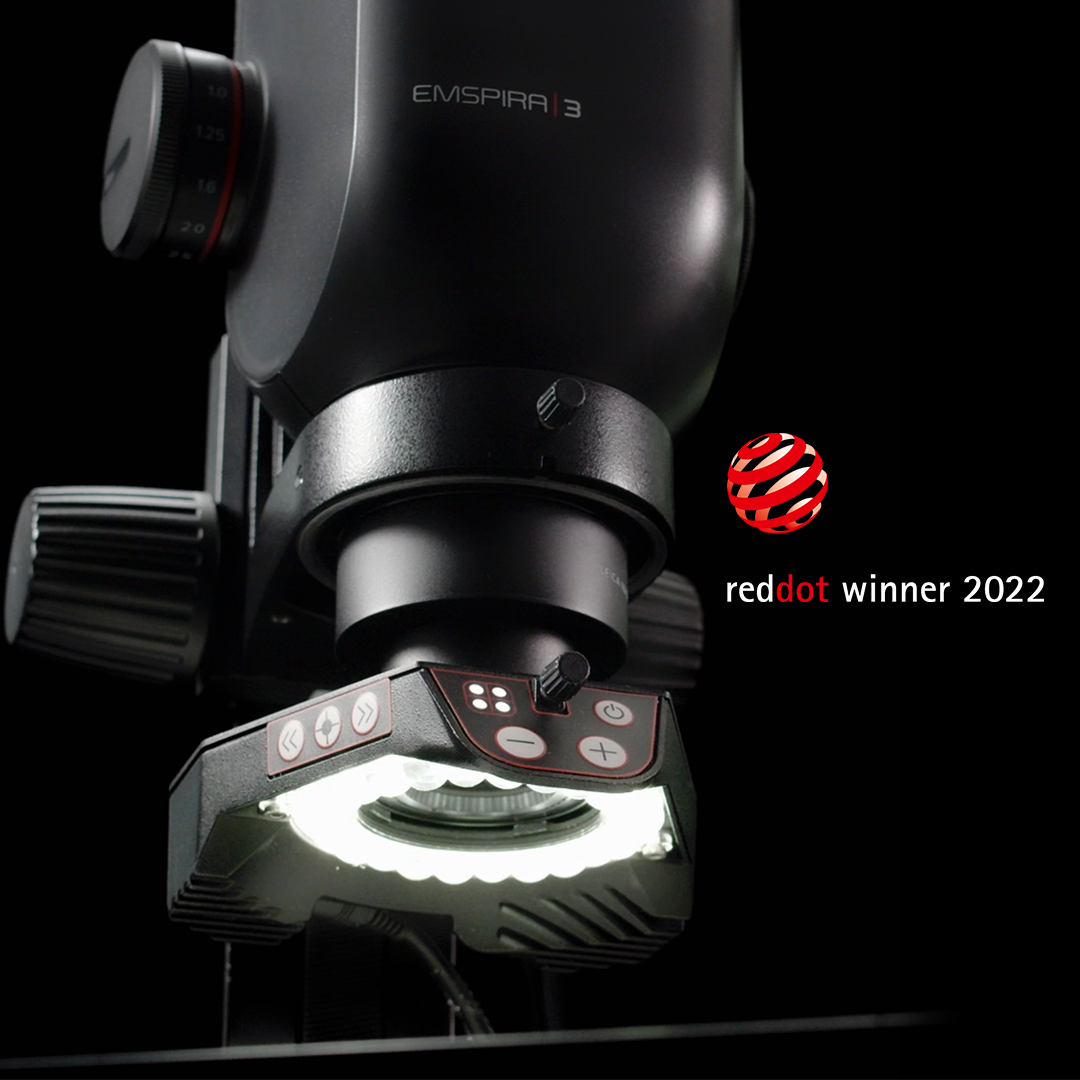
Emspira 3 digital microscope – inspiring simple inspection
The Emspira 3 digital microscope empowers users to streamline inspection processes, have easy access to images, and provides them a flexible solution they can rely on. As the winner of a 2022 Red Dot Award for product design, the system has an innovative modular design and a wide range of accessories and illumination options. Emspira 3 combines everything needed to perform comprehensive visual inspection into a single system, including comparison, measurement, and documentation sharing.
2020


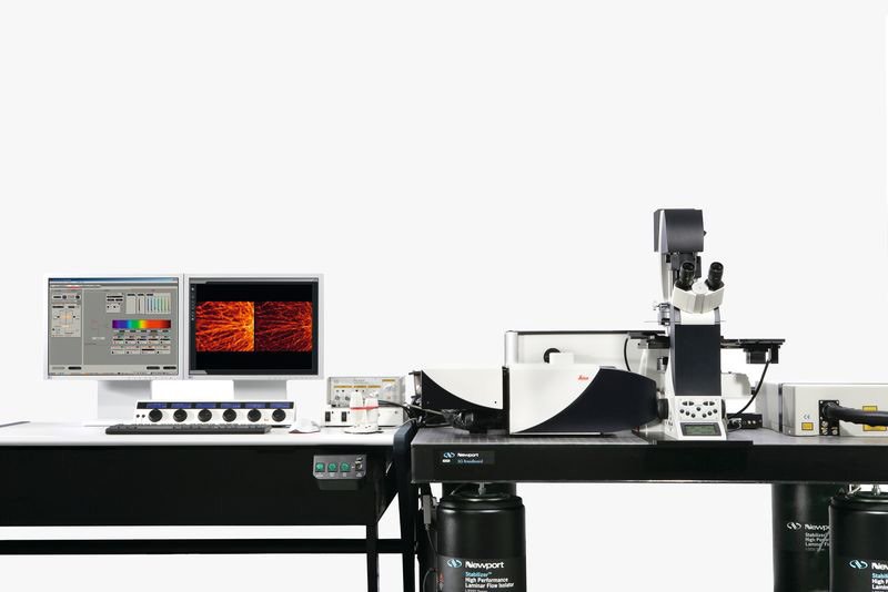
Confocal imaging that gets you closer to the truth.
STELLARIS is a completely re-imagined confocal microscope platform that can be combined with all Leica modalities, including FLIM, STED, DLS, and CRS. It gives researchers the power to see more and, as a result, collect more accurate and reliable data to prove hypotheses with precision. STELLARIS gives access to lifetime-based information in every experiment, in parallel with common intensity imaging approaches. With innovative technologies such as White Light Lasers (WLL), TauSense imaging tools and the Image Compass user interface, scientists have the power to see more, the potential to discover more and the productivity to do more.
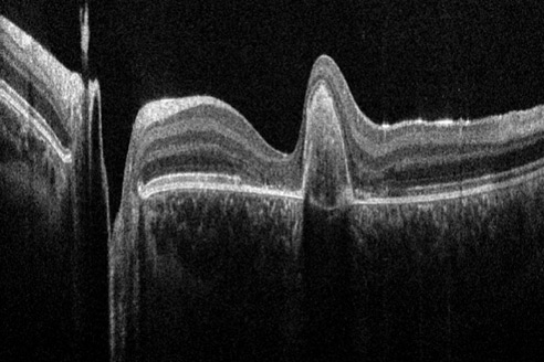
Intraoperative OCT imaging system EnFocus
EnFocus intraoperative optical coherence tomography (OCT) allows ophthalmic surgeons to see what lies underneath the surface. It provides additional real-time information for a deeper understanding of how subsurface tissue reacts to surgical maneuvers during eye surgery. EnFocus built into the Proveo 8 ophthalmic microscope supports workflow enabling surgeons to independently control the OCT view via footswitch, handle or touchscreen.
2019
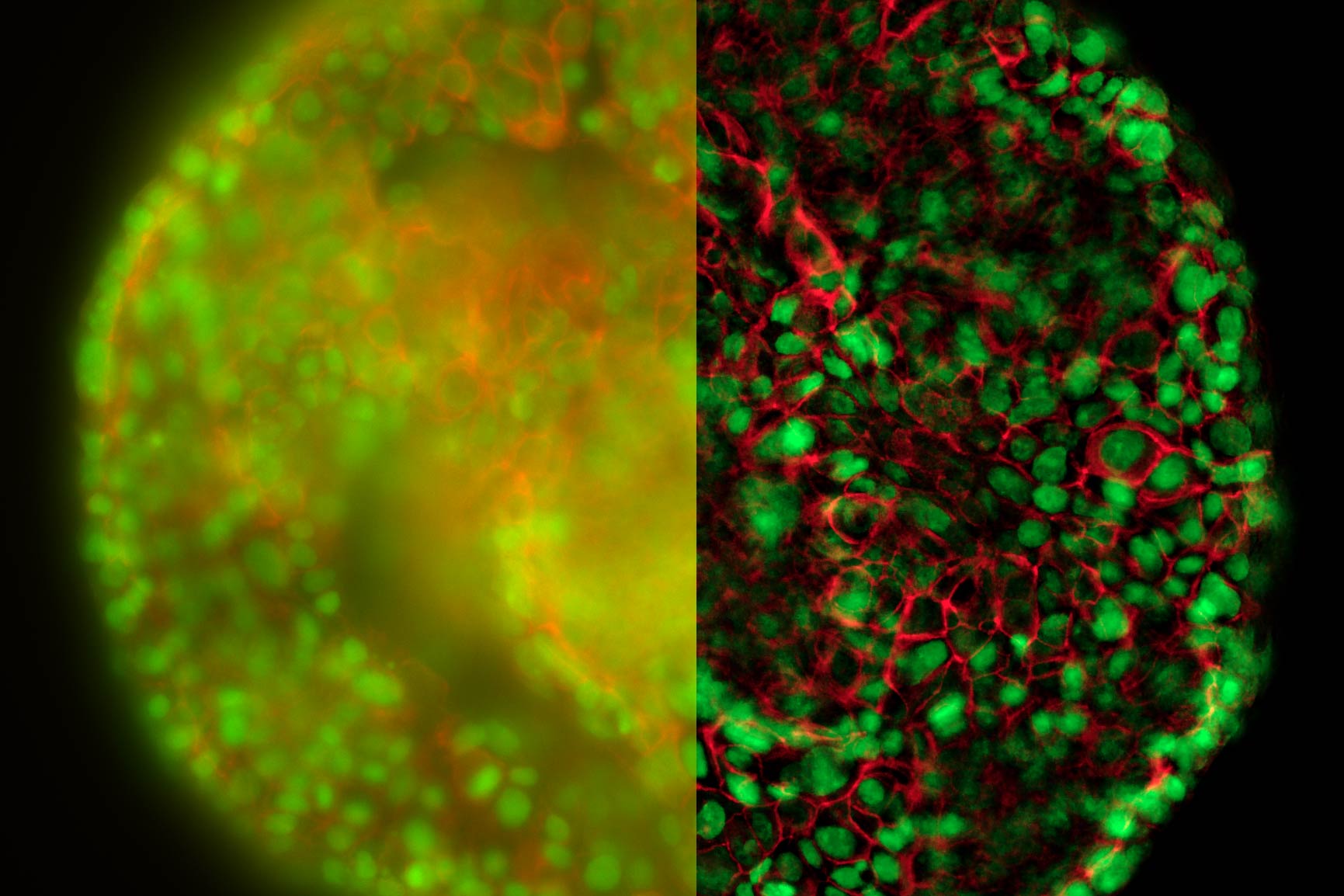
Widefield Visualization of 3D Biologically Relevant Specimens Transformed
The THUNDER Imagers enable users to visualize clearly in real time fine details even deep inside thick specimens of biologically relevant models, e.g., model organisms, tissue sections, and 3D cell cultures. The Imagers eliminate the out-of-focus haze that clouds the view of thick specimens when using camera-based fluorescence microscopes. This performance advantage is achieved with a new opto-digital method created by Leica Microsystems called Computational Clearing.
2018
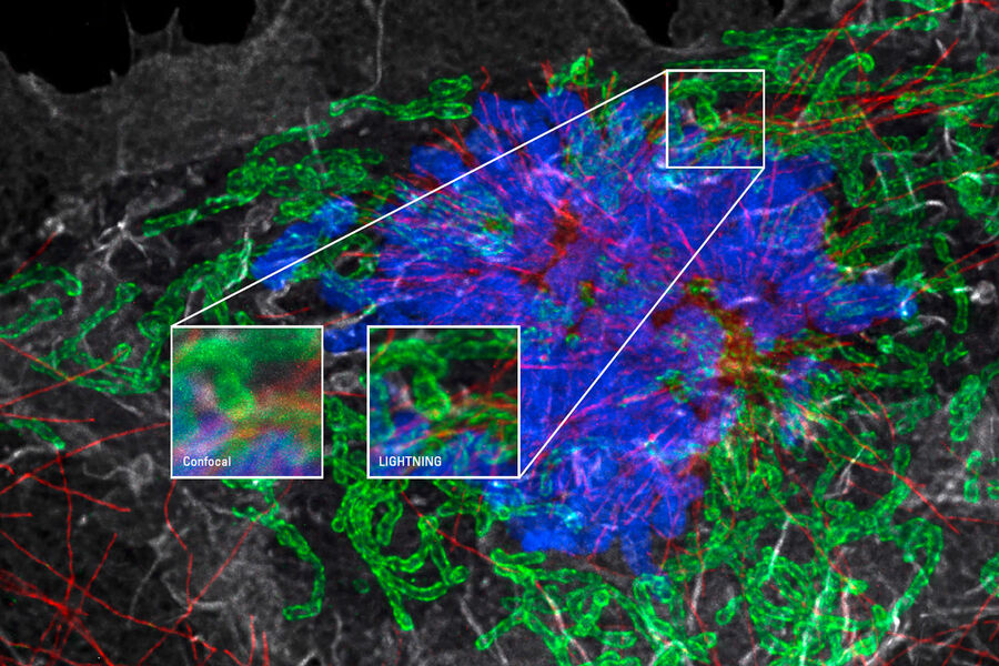
Redefining the Detection Limit for Confocal Imaging
LIGHTNING extracts valuable image information from previously unseen or inaccessible fine structures and details, expanding imaging capabilities both in the classic confocal range and beyond the diffraction limit, down to 120 nm.
Lifetime imaging in an instant
The SP8 FALCON (FAst Lifetime CONtrast) system can record lifetime contrast 10x faster than previous solutions. This lifetime information lets researchers monitor interactions between proteins in living cells. Additionally, it provides extra contrast to clearly separate a multitude of fluorophores.
Augmenting intraoperative reality
The ARveo digital augmented reality neurosurgical microscope provides surgeons with extensive visual information to empower their intraoperative decision-making. The microscope features groundbreaking GLOW AR digital technology, which combines white light with fluorescence for a single, precise view of the surgical site. GLOW800 AR fluorescence, the first AR modality powered by GLOW AR technology is available fully integrated into ARveo. In combination with ICG, GLOW800 allows surgeons to observe cerebral anatomy in natural color, augmented by a real-time view of blood flow. This is particularly useful in AVM or aneurysm surgeries. The ARveo also integrates digital imaging from IGS systems and endoscopes, as well as allowing surgeons to work “heads-up” looking at a large 4K 3D monitor.
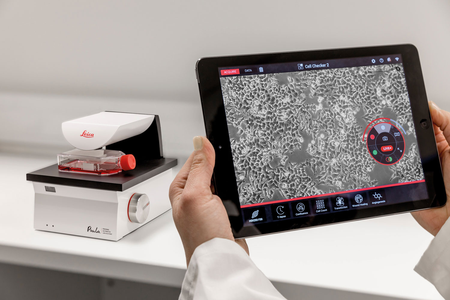
Routine tasks in the cell culture lab become digitized
PAULA (Personal AUtomated Lab Assistant) helps to speed up daily cell culture tasks and standardize the results to improve the downstream workflow. It is configured to stay by your cells on any lab bench or even directly in an incubator.
Get Quality Serial Sections for Array Tomography Fast
ARTOS 3D marks a new level of quality and speed for ultramicrotome sectioning. ARTOS 3D automatically produces ribbons of extremely consistent, thin sections (nanometer range thickness) for array tomographic 3D image reconstruction of the specimen.
Helping surgeons see more, simply
With the launch of the PROvido multidisciplinary microscope Leica Microsystems enhances intraoperative visualization in a broad range of surgical applications. PROvido features exclusive FusionOptics technology from Leica Microsystems, which was previously only available in premium microscope solutions. Surgeons can now see a larger area in full focus and benefit from less interruptions through time consuming refocusing. The top-of-the-line optics and illumination combined for the first time in a responsive, stable floor stand, allow surgeons to see more, simply.
2017
The world‘s first tunable deep imaging solution
With the launch of the all new SP8 DIVE system Leica Microsystems offers the world’s first spectrally tunable solution for multi-color, multiphoton deep tissue imaging. Equipped with 4Tune, a tunable, non-descanned detection system, the SP8 DIVE offers you unlimited flexibility and enables you to develop new multicolor deep in vivo experiments. DIVE stands for Deep In Vivo Explorer and enables researchers to capture up to four fluorophores simultaneously and an unlimited number of fluorophores sequentially. The ability to visualize complex biological processes in living tissues over time is key for driving research in the fields of neurosciences, cancer and inflammatory diseases.
Find, observe and interact with living cells like never before
Whether you need to precisely follow the development of a single cell in a dish, screen through multiple assays, obtain single molecule resolution, or tease out behaviors of complex processes, a DMi8 S system will enable you to see more, see faster, and find the hidden to find the missing link. The DMi8 S imaging solution from Leica provides 5x more speed, and an increased viewing area up to 10,000x. This can be combined with the new photomanipulation scanner to activate, ablate, and bleach within one experiment. For super resolution and nanoscopy, the Infinity TIRF has been added allowing simultaneous multi-color imaging with single molecule resolution. Opening up the next chapter in widefield imaging.
2016
Preparing the ground for future innovations:
Leica Microsystems exclusively licensed SCAPE microscopy for Life Science applications from Columbia University, a technique for fast 3D imaging of living samples with a sheet of light – which complements own IP and developments as well as its exclusive license of Oblique Plane Microscopy (OPM) from Imperial College.
Worlddidac Award for Leica EZ4 W educational stereo microscope:
The Leica EZ4 W which transfers HD images directly to students' mobile devices has been recognized with the prestigious Worlddidac Award, an award that honors outstandingly innovative educational products or solutions.
New image injection technology guides surgeons:
CaptiView technology injects images from image-guided surgery (IGS) software into the microscope eyepieces. Surgeons can fully concentrate on their patient, have all the data in front of their eyes and do not need to look up during the procedure.
Leica Optical Coherence Tomography (OCT) systems support ophthalmologists, ophthalmic surgeons, and researchers with easy-to-use, high-quality imaging technology.

2015
Helping researchers understand life-sustaining processes:
The first High Pressure Freezer with integrated light stimulation is a technology so precise that researchers can start asking new questions – and will be able to get answers.
Leica Microsystems acquires Optical Coherence Tomography (OCT) company Bioptigen:
The contact-free imaging method aids ophthalmologists and researchers in the diagnosis of physiological and pathologic conditions of the eye.
Wi-Fi-capable microscopy instruments help increase student learning time:
The Leica EZ4 W educational stereo microscope with built-in wireless camera and the Leica ICC50 W digital camera transfer HD images directly to students' mobile devices.
2014
Nobel Prize for Stefan Hell, father of super-resolution microscopy:
Stefan Hell was awarded the Nobel Prize in Chemistry for the development of super-resolved fluorescence microscopy. In collaboration with Leica Microsystems, this principle was translated into the first commercially available STED microscope.
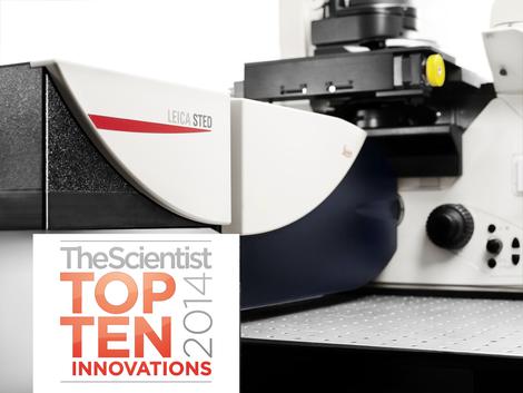
Two awards for the Leica TCS SP8 STED 3X:
The Scientist Top 10 Innovations Award and R&D 100 Award recognize the super-resolution microscope as one of the innovations that will change the way life scientists work.
Supporting surgeons in visualization:
The surgical microscope Leica M530 OH6 is equipped with technology to enable surgeons to see better into deep, narrow cavities.
Building-block concept for inverted research microscope:
Researchers can build their Leica DMi8 microscope fully modularly. The platform can be adapted to their needs any time – a big difference to former instruments.
Microscopy in Outer Space:
Astronaut Koichi Wakata of the Japan Aerospace Exploration Agency used the inverted research microscope Leica DMI6000 B to carry out live cell experiments at the International Space Station.
2013
Award for Leica SR GSD 3D super-resolution microscope
Enabling scientists to visualize and study cellular structures and processes down to the molecular level, the instrument was voted among the Top 10 Innovations 2013 for laboratories and research.
Integrated 3D visualization for neurosurgeons:
First surgical microscopes with integrated TrueVision 3D technology enable an entire operating team to see what the surgeon sees without the need for a separate cart, thus saving space in the operating room.
Leica Biosystems and Leica Microsystems strengthen market position in Brazil:
Acquisition of Aotec, distributor for more than 25 years moves presence in Latin America forward.
2012
2012 MX Award for Leica Microsystems headquarters:
For its switchover to Kanban systems, Leica Microsystems Operations in Wetzlar, Germany, won the Manufacturing Excellence award for logistics and operational management.
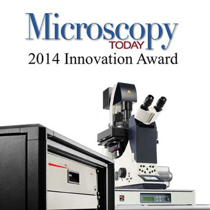
Three awards for Leica GSD super-resolution microscope:
R&D magazine with its R&D100 awards for excellence in technological innovation, one of the three related "Editor's Choice Awards", and the Top Ten Innovations Awards 2012 by US American magazine Microscopy Today.
Enabling new discoveries:
The Leica TCS SP8 confocal microscope combines high-performance optics, the fastest true confocal scanner and the most sensitive detection system available, to enable researchers to uncover life's secrets.
The Leica Science Lab hosts articles, interviews, tutorials, and webinars for users.
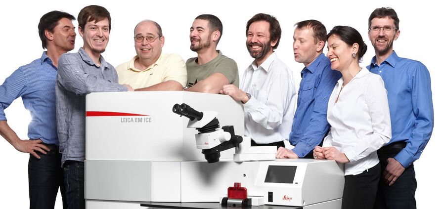
Leica EM ICE team who had more than 145 years of experience in EM sample preparation when they launched the Leica EM ICE
2011
Learn, share, contribute. Science Lab goes live:
Science Lab is the knowledge portal on microscopy and EM sample preparation. Articles, interviews, tutorials, and webinars help users with interesting and hands-on information. By 2016, Science Lab has grown to an online resource with more than 600 articles.
2011 MX Award for Leica Biosystems Nussloch:
Leica Biosystems scores in the customer focus category of the Manufacturing Excellence (MX) Award 2011.
Classroom in HD:
Students and teachers can now view microscope images in HD quality during microscopy classes which enables them to detect more detail in the images.

2010
Award for Remote Care Service Concept:
Leica Microsystems received a gold M2M Value Chain award with Axeda Corporation honored as its enabler at the annual Connected World Conference.
Strategic Partnership:
Kavo Dental and Leica Microsystems cooperate for dental microscopes.
Frost & Sullivan Tissue Diagnostics award:
Leica Biosystems receives the North American Product Strategy Award in Tissue Diagnostics by research and consulting firm Frost & Sullivan.
2009
Exclusive license for a new generation of optical microscopes:
Max Planck Innovation grants exclusive license for the new GSDIM (ground state depletion microscopy followed by individual molecule return) super-resolution method to Leica Microsystems.
Slide scanner for optimal histological examinations:
The Leica SCN400 Slide Scanner offers an alternative to the microscope for the examination of histological samples in pathology, research, and teaching.
Quantitative particle analysis:
The Leica Cleanliness Expert is an all-in-one system for measuring impurities in cleaning liquids for micro-mechanic and motor components during production in the automotive industry. This helps failure analysis and quality assurance.
2008
Center of Excellence:
Leica Microsystems becomes a founder partner of the European Molecular Biology Laboratory (EMBL) Advanced Training Centre in Heidelberg, Germany.
Award for the Leica White Confocal:
The Scientist Top 10 Innovations Award 2008 for the Leica TCS SP5 X Supercontinuum Confocal Microscope.
Award for innovation in stereo microscopy:
Leica Microsystems wins PRODEX AWARD for FusionOptics, a technology that creates images with high resolution and a greater depth of field for better 3D impression.
Seeing better and more in neurosurgery:
Leica M720 OH5 - the world's most compact neurosurgical microscope with horizontal optics with patented mobility concept and greatest overhead maneuverability.
2007
Breaking the diffraction barrier:
With the super-resolution microscope Leica TCS STED light microscopy goes beyond the limits. This first commercial STED microscope achieves an optical resolution of less than 90 nm.
Step beyond infinity:
With the high-end stereo microscopes Leica M165 C and M205 C with FusionOptics, Leica Microsystems sets a new standard in stereo microscopy.
Leica Microsystems' new Biosystems Division:
After integrating Vision BioSystems, the Leica Biosystems division was formed – as the only source to offer instruments for the entire histo-pathology process.
Three new products for EM sample preparation:
Leica EM TRIM2 specimen trimmer, the Leica EM TXP for cutting, milling, and polishing, and the Leica EM RAPID milling system.
Licensing Agreements:
Max Planck Innovation concludes licensing agreement for RESOLFT technology; Harvard University's Office of Technology Development licensed its CARS microscopy technology to Leica.

2006
Network solution for histopathologists:
The Leica DMD108 increases physical comfort, significantly speeds up daily workflow without changing the process, and provides an easy-to-use network solution for sharing data.
Third "Innovationspreis" (German Business Innovation Award) for Leica Microsystems:
The company wins the award in the medium-sized business category. The company already won the prestigious award in 1984 for the ELSAM acoustic microscope and again in 2002 for the DUV high-resolution microscope objective for photomask and wafer fabrication.
2005
Innovative laser microdissection system:
With its enhanced laser technology, the Leica LMD6000 is suitable for processing thicker specimens and harder materials, making it ideal also for botanical research applications.
The only broadband confocal:
The Leica TCS SP5 features the broadest band of imaging speeds and resolutions ever available in one Confocal Microscope.
Integrated live cell workstation:
The Leica AF6000 LX integrated system for advanced widefield fluorescence imaging and analysis enables researchers to study the processes of life by imaging fast cell dynamics or 4D experiments over several days.

2004 - 1847
2004: First Super-Resolution Confocal (4Pi) Microscope
2003: German Business Innovation Award for the Leica DUV objective
1998: The Business Divisions of the Leica Group, Leica Camera, Leica Microsystems and Leica Geosystems, become three independent companies
1993: 1st Joint Venture Opening of the Leica group in China for specimen preparation.
1990: Merger of Wild Leitz, Cambridge Instruments, Reichert & Jung and Bausch & Lomb to form the Leica Group
1986: Merger of Ernst Leitz and Wild Heerbrugg to form the Wild Leitz Group
1984: German Business Innovation Award for the ELSAM acoustic microscope
1981: Foundation of the Wild Leitz group.
1976: Metals Research expanded and bought Cambridge Instruments (1st manufacturer of Scanning Electron Microscopes).
1972: Beginning of cooperation between Leitz Wetzlar and Wild Heerbrugg.
1967: Image Analysis (Quantitative Microscopy) brought to market
1932: Incident Light Fluorescence Microscopy introduced
1929: Photo Microscopy brought to market
1925: First Polarizing Microscope introduced
1921: Foundation of the optical company Wild Heerbrugg in Switzerland.
1914: Leitz’ small-format 35 mm camera (‘Leica’) invented by Oskar Barnack
1913: First Binocular Microscope presented
1907: The 100.000th microscope is presented to Nobel prize winner Robert Koch.
1881: Charles Darwin’s son Horace founds the optical company Cambridge Instruments
1876: Optical company C. Reichert founded in Vienna
1872: Precision Engineering company R. Jung founded in Heidelberg
1869: Ernst Leitz takes over "Optical Institute" and changes the company name into “Ernst Leitz”
1853: Foundation of the Bausch & Lomb Instruments Division in USA.
1849: Carl Kellner’s Optical Institute founded in Wetzlar
1847: Foundation of Spencer Lens/American Optical Instruments in USA.
