Light Sheet Microscopes
Light Sheet Microscopes

When should I use light sheet microscopy?
Use light sheet microscopy when you need fast 3D imaging, reduced phototoxicity, and long-term observation. The method illuminates only the focal plane. This makes Leica light sheet microscopes particularly suitable for live imaging, volumetric acquisition, and dynamic biological processes.

How to reduce phototoxicity in live imaging?
Light sheet microscopy restricts illumination to the focal plane, which lowers total light dose and limits photobleaching. Leica light sheet microscopes, such as Viventis SCAPE, Viventis Deep, and STELLARIS DLS, apply this approach to enable gentle live imaging and longer observation of sensitive biological processes.

How can I image organoids in 3D?
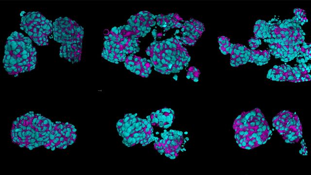
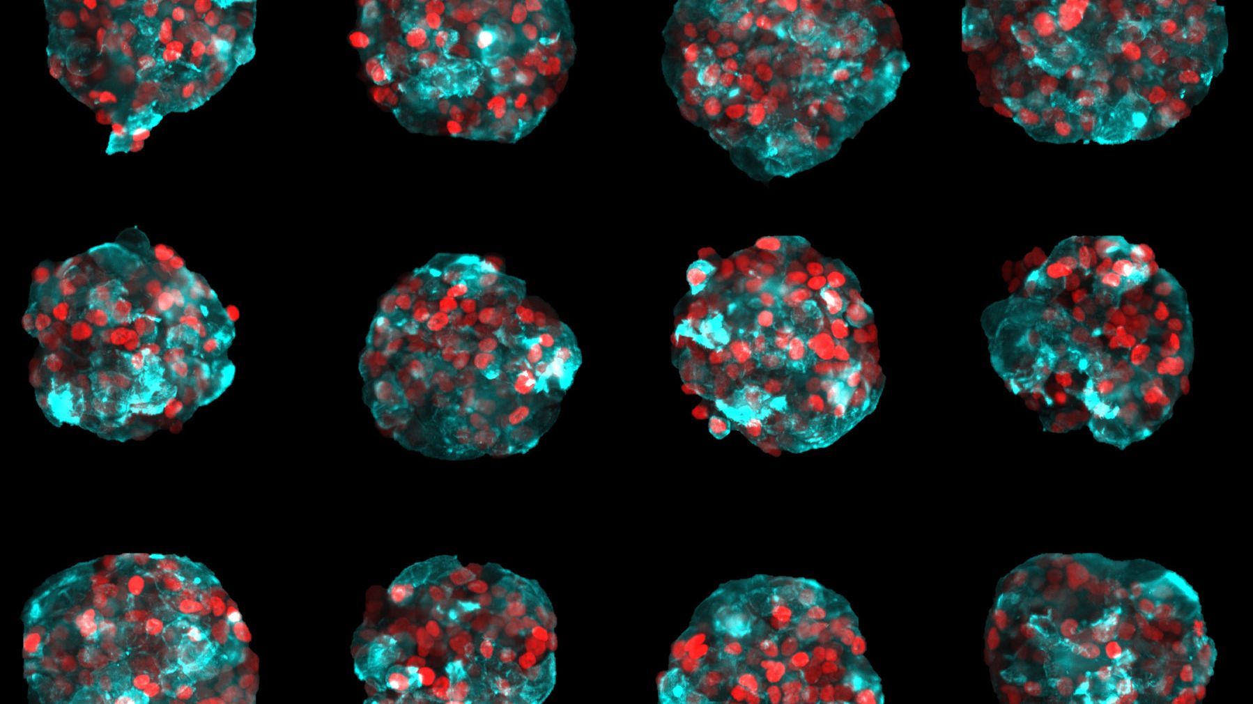
Yes, light sheet microscopy is well suited to organoids and 3D cell culture models because it delivers fast volumetric imaging with low light exposure, supporting longer live observations. Leica offers systems ranging from dual-view setups for deep imaging in scattering samples to single-objective designs compatible with microplates and slides.

How do I image cleared samples?
Leica offers switchable optics on the Viventis Deep system to adapt to cleared specimens and capture organ volumes while preserving structural context. You can image large samples in 3D with light sheet microscopy combined with tissue clearing. Refractive index matching reduces scattering for deeper, higher-contrast imaging.
What applications benefit from Leica light sheet microscopes?
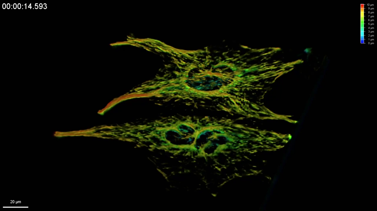
Fast and gentle volumetric imaging
Fast and gentle imaging is essential to capture dynamic biological processes without compromising sample viability. Viventis SCAPE, using OPM/SCAPE technology, enables fast, gentle imaging in standard sample carriers without the need for special sample preparation. Viventis Deep, with dual illumination and dual detection, enables deep imaging of larger, more light scattering samples while maintaining good resolution.
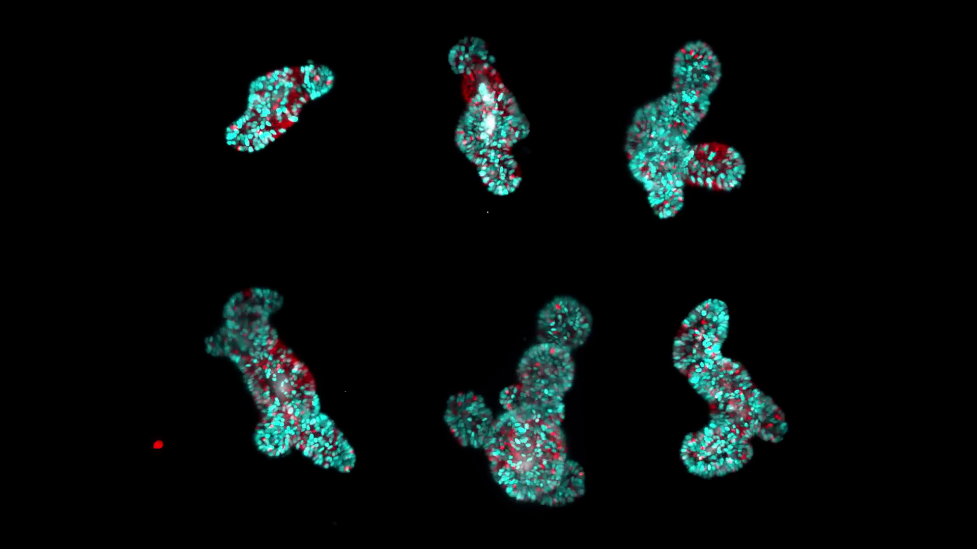
Time-lapse imaging
Time lapse imaging is a cornerstone of developmental biology, stem cell research, and cancer studies. Leica’s light sheet microscopes protect subtle physiological signals and provide stable culture conditions with easy sample access – supporting long-term experiments over days and weeks.
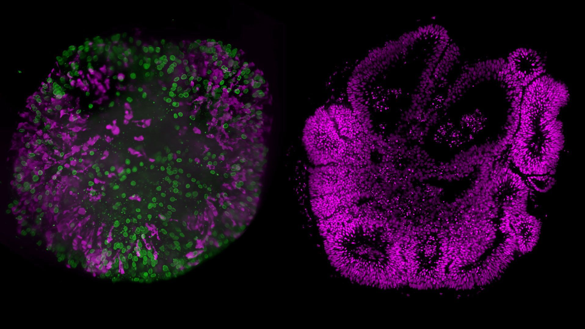
Cleared sample imaging
Clearing removes light scattering, making large, thick tissues optically transparent and accessible deep inside the sample. Cleared sample imaging with Viventis Deep allow high-resolution, high-contrast 3D imaging for imaging samples ranging from hundreds of micrometers up to 9x6x6 mm in size.
Typical applications covered by Leica light sheet microscopes
Viventis SCAPE, Viventis Deep and STELLARIS DLS cover a wide range of applications from live and fixed imaging to cleared samples. The sample sizes range from approx. 50 μm to 2 mm and to volume sizes of 6x6x9 mm for cleared imaging.
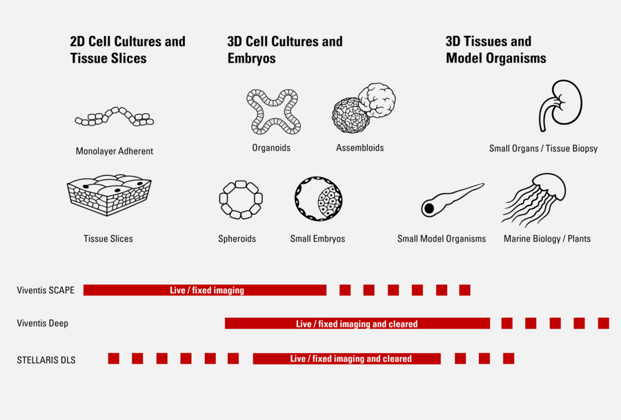
Our Light Sheet Microscope Solutions
Viventis SCAPE | Viventis Deep | STELLARIS DLS | |
| Configuration | Inverted with single-objective for both illumination and detection | Inverted with dual illumination and dual detection | Upright with dual illumination and single detection on a confocal platform |
| Sample mounting | Standard imaging dishes up to multi-well plate | Dedicated open top multi-well sample holder | Custom mounting into standard imaging dishes. Dipping objectives |
| Sample type imaging | Live imaging Fixed sample imaging | Live imaging Fixed sample imaging Cleared sample imaging | Live imaging Fixed sample imaging Cleared sample imaging |
| Acquisition speed | xxx | x | x |
| Imaging in depth | x | xxx | xx |
| Multimodality /advanced features | Simultaneous multi-color imaging | Photomanipulation | Confocal, Photomanipulation |
| Go to product page | Go to product page | Go to product page |
Frequently Asked Questions Light Sheet Microscopes
Light sheet microscopy is an imaging technique that uses a thin sheet of light to illuminate a sample from the side. A camera or detector captures the emitted fluorescence. It allows for rapid, high-resolution imaging of living volumetric specimens with reduced phototoxicity and photobleaching.
Light sheet microscopy confines illumination to a thin optical section rather than the entire sample volume. This substantially reduces unnecessary light exposure, helping preserve biological integrity, maintains physiological conditions, and enable extended imaging of delicate or light sensitive specimens.
Light sheet imaging is well suited for thick, delicate or dynamic samples, including live embryos, organoids, spheroids, 3D cell cultures, fixed tissues, and optically cleared specimens.
Yes. Light sheet microscopy is commonly referred to as light sheet fluorescence microscopy (LSFM) or selective plane illumination microscopy (SPIM). These terms describe closely related implementations that share the same core principle of planar illumination combined with widefield detection.
Yes. By illuminating only the focal plane, light sheet microscopy minimizes phototoxicity and photobleaching. This makes it well suited for live imaging experiments, including long term time lapse studies of sensitive biological processes in living samples.
Imaging cleared samples is ideal when large or thick specimens need to be visualized in 3D with full depth access. Clearing methods reduce light scattering due to refractive index matching within the sample, enabling high-resolution, high contrast imaging deep inside tissues. It is especially useful for samples, where capturing complete spatial context and analysis of tissue architecture is desired. It allows structures to be analyzed across entire organs while combining organ level overview with cellular or even subcellular detail in one dataset.