Digital Microscopes

Digital Microscopes

Digital Microscopes

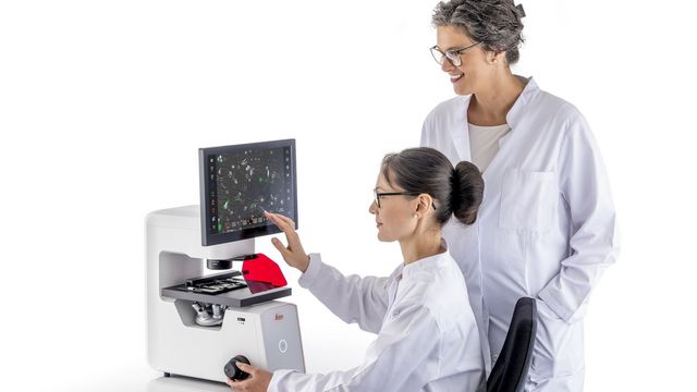
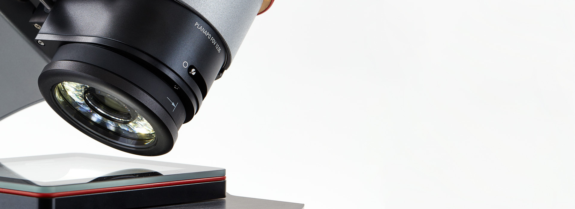
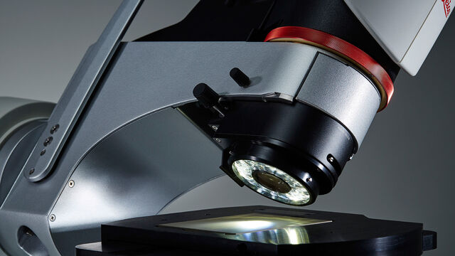
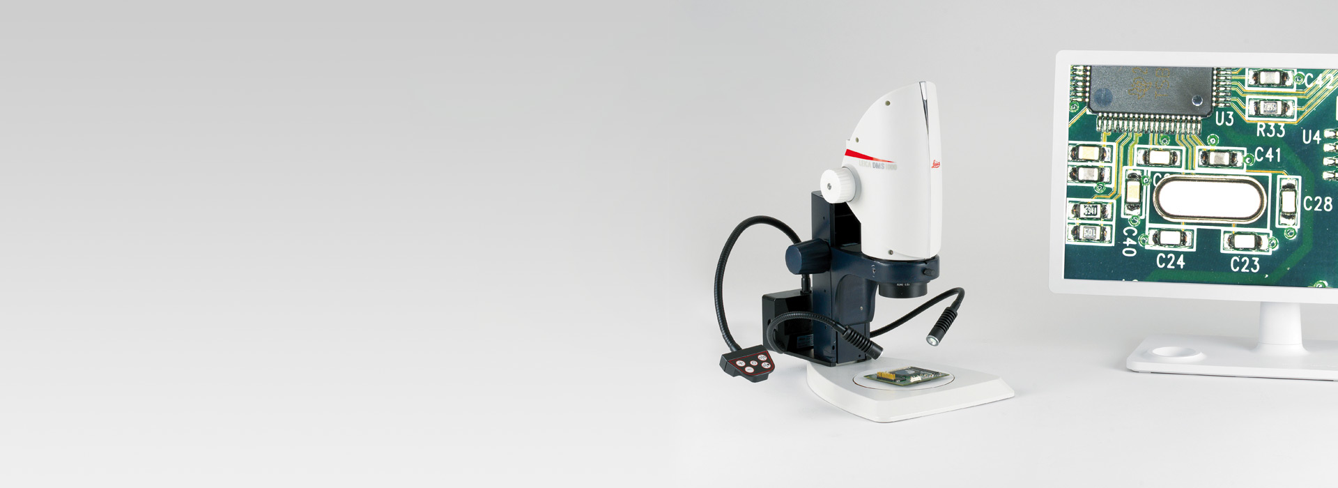
Digital microscopes are microscopes without eyepieces. A digital camera acts as a detector. Images are displayed on a screen or monitor, turning the microscopy workstation into an ergonomic digital workplace.

Digital Microscopes

Digital microscopes for inspection, quality control, and failure analysis as well as 2D and 3D measurement and surface metrology. By Leica Microsystems.

Read our latest articles about Digital Microscopes

The knowledge portal of Leica Microsystems offers scientific research and teaching material on the subjects of microscopy. The content is designed to support beginners, experienced practitioners and scientists alike in their everyday work and experiments.
More Articles

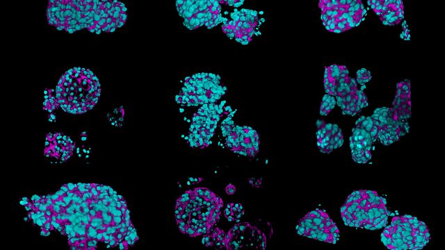
What’s the Best Organoid Imaging Approach for Early Drug Discovery?
Organoids and other complex in vitro models (CIVMs) are becoming increasingly important in early drug discovery and translational research, driven by the need for more predictive, human-relevant data in line with evolving regulatory expectations. However, scientists face many challenges when imaging and analyzing these 3D systems. This eBook explores how complementary imaging and analysis workflows can help you extract deeper insights from complex 3D biological models.

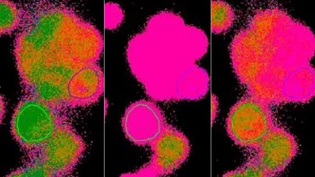
Ratiometric Imaging and Analysis of Ion Concentration in Cells
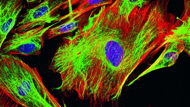
Many cellular functions depend on the dynamic balance of ions, electric potentials, and pH between the cytosol and surrounding extracellular space. Changes in these values affect cellular function. Measuring cellular ion, potential, and pH dynamics is very interesting for biology research, but is difficult to do with conventional fluorescence. How ratiometric imaging overcomes this limitation by allowing the observation of changes in fluorophore emission wavelengths and intensities is explained.

Overview of Fluorescent Dyes in terms of Applications and Properties
An introduction to commonly used fluorescent dyes and an overview of their characteristics are given in this article. Fluorescence microscopy is used for the study of specific cellular components with the help of fluorescent dyes, fluorescent proteins, or immunofluorescent stains using antibodies. Thanks to a large variety of fluorescent agents, fluorescence microscopy can be used to examine proteins, nucleic acids, glycans, organelles, and other cellular structures.

How to Select the Right Measurement Microscope
With a measurement microscope, users can measure the size and dimensions of sample features in both 2D and 3D, something crucial for inspection, QC, failure analysis, and R&D. However, choosing the right microscope involves assessing application needs along with microscope performance, ease of use, and flexibility. Today, measurements are often made digitally, i.e., a microscope with a camera and software and images displayed on a monitor, rather than via an eyepiece reticule, leading to increased precision and repeatability. Analyze samples reliably and quickly with the right measurement microscope.

Microscope Calibration for Measurements: Why and How You Should Do It
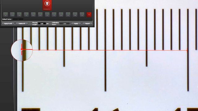
Microscope calibration ensures accurate and consistent measurements for inspection, quality control (QC), failure analysis, and research and development (R&D). Calibration steps are described in this article. Using a reference, calibration enables reproducible results and helps ensure agreement with guidelines and standards. To attain accurate and consistent results, it is recommended to calibrate a microscope and regularly check it. If needed, support can be obtained from calibration experts.

Burr Detection During Battery Manufacturing
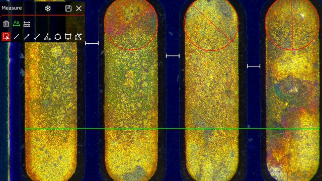
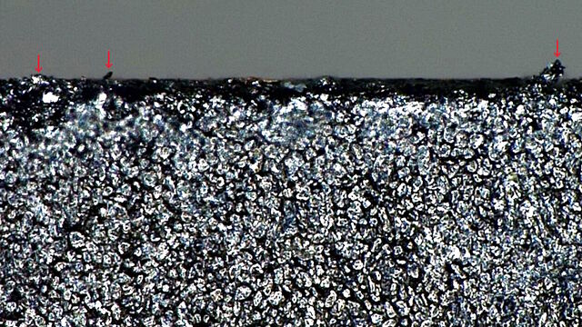
See how optical microscopy can be used for burr detection on battery electrodes and determination of damage potential to achieve rapid and reliable quality control during battery manufacturing.

Guide to Live-Cell Imaging

For a wide range of applications in various research fields of life science, live-cell imaging is an indispensable tool for visualizing cells in a state as close to in vivo, i.e. living and active, as possible. This guide reviews a wide range of important considerations for ensuring successful live-cell imaging and introduces a variety of high-performance solutions designed to overcome common challenges. These advances enable new insights into cellular physiology and dynamics.

Microscopy and AI Solutions for 2D Cell Culture
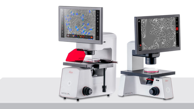
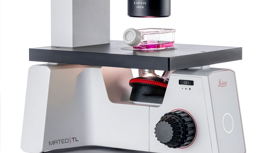
This eBook explores the integration of microscopy and AI technologies in 2D cell culture workflows. It highlights how traditional imaging methods—such as brightfield, phase contrast, and fluorescence—support routine cell monitoring, while digital platforms like the Mateo TL and FL enhance reproducibility through automated confluency checks, cell counting, and transfection analysis. It also shows how integrated data management, audit trails, and sample tracking improve documentation and research integrity. The book concludes with a look at future trends, including microfluidics and hybrid 2D–3D systems, emphasizing the growing need for advanced imaging tools to meet evolving research demands.

A Microvascular Surgeon’s View: How MyVeo Transforms Visualization
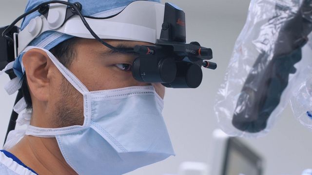
In this article, Dr. Andrew T. Huang, MD, FACS, otolaryngologist and a head and neck reconstructive surgeon, shares how digital 3D surgical visualization with the MyVeo headset from Leica Microsystems transforms his practice. For microvascular and nerve repair procedures, he discusses how it helps him focus, train and collaborate with his OR team in a comfortable and relaxed position during surgery. The surgical visualization headset also seamlessly integrates into the OR. Learn how digital 3D viewing technology and augmented reality visualization are shaping the future of surgery.

How to Image Axon Regeneration in Deep Muscle Tissue

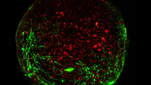
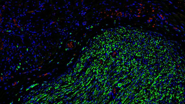
This study highlights Dr. Aaron Lee’s research on mapping nerve regeneration in muscle grafts post-amputation. Limb loss often leads to reduced quality of life, not only from tissue loss but also due to neuropathic pain caused by disorganized axon regrowth. Histological and fluorescence imaging on Mica can provide complementary insights into axon outgrowth and branching during nerve regeneration. This research could help shape future neuroprosthetic interface designs, improving patient outcomes.

A Guide to Neuroscience Research

Neuroscience often requires investigating challenging specimens to better understand the nervous system and disorders. Leica microscopes helps neuroscientists obtain insights into neuronal functions.

The Guide to Augmented Reality in Microsurgery

In an era of technological advancement, Augmented Reality (AR) is rapidly transforming the medical field. In surgical microscopy, AR can display fluorescence signals as digital overlays in real-time within the surgical field of view, providing surgeons with additional information about fluorescent areas of interest. A surgeon's view can be augmented with data overlays from different systems, offering functional or anatomical information superimposed onto the live microscope view.

Automotive Part Verification and Development according to Specifications

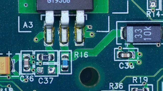
Automotive part verification during the development and production of parts and components by suppliers or manufacturers is important for ensuring that specifications are met. Specifications are critical for maintaining performance standards and safe operation of vehicles. However, there is always demand for efficient and cost-effective R&D and production while meeting ever-stricter quality standards. This article shows how investigation and documentation of auto parts and components to verify specifications can be done easily and quickly with digital microscopy.

Mica: A Game-changer for Collaborative Research at Imperial College London
This interview highlights the transformative impact of Mica at Imperial College London. Scientists explain how Mica has been a game-changer, expanding research possibilities and facilitating interdisciplinary collaboration. They explain how detailed live cell imaging with Mica provides more meaningful information, keeping scientists at the forefront of research. The team foresees Mica continuing to open new research avenues, including the study of microfluidics and other advanced applications.

Depth of Field in Microscope Images

For microscopy imaging, depth of field is an important parameter when needing sharp images of sample areas with structures having significant changes in depth. In practice, depth of field is determined by the correlation between numerical aperture, resolution, and magnification. For the best possible visualization of samples, modern microscopes can be adjusted to produce an optimum balance between depth of field and resolution. In theory, these parameters are inversely correlated.

Introduction to 21 CFR Part 11 for Electronic Records of Cell Culture

This article provides an introduction to the recommendations of 21 CFR Part 11 from the FDA, specifically focusing on the audit trail and user management in the context of cell-culture laboratories. It is intended for professionals in the biotechnology and pharmaceutical industries who are responsible for ensuring agreement with 21 CFR part 11 for electronic records and electronic signatures. A digital microscope approach, e.g. with Mateo FL, offers the advantage of more consistent and efficient electronic documentation of cell-culture results compared to a paper-based method.

Key Questions

1Applications for Digital Microscopes?

Digital microscopes are ideal instruments for analysis and documentation of parts and samples during research and development (R&D), manufacturing and inspection, quality control and assurance (QC/QA), as well as failure analysis (FA).

2Advantages of digital microscopes?

The most obvious advantage is the ergonomics of the instrument followed closely by ease-of-use for routine work.

3What does "encoding" mean?

A microscope is "encoded" when its hardware is in direct communication with computer software and when specific parameter values are tracked and saved with the image data. These specific parameters are defined and, therefore, referred to as encoded parameter values.
Leica Digital Microscope Setup

Inspection, documentation, and digital analysis without eyepieces

Leica Digital Microscopes have become increasingly popular in production, quality control and quality assurance, failure analysis, research and development, as well as forensics for their excellence in inspection, documentation, analysis in both 2D and 3D.

Constant customer feedback allows our product management and R&D to continue to bring you instruments that both meet your needs and exceed your expectations throughout your working day.

Why use Leica digital microscopes?

Leica Microsystems take great pride in being able to deliver digital microscopes that are optimized over the entire inspection, documentation, and analysis workflow.

Easy operation

Leica digital microscopes can be handled intuitively over the entire microscopy workflow. One example: This video shows how an objective can be exchanged one-handedly.

Smooth workflow

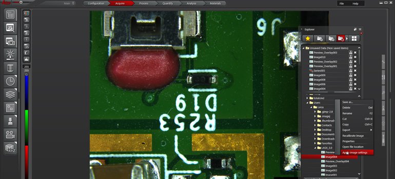
Leica digital microscopes streamline the workflow from positioning the sample to generating a report. Software additionally speeds up handling.

Perfect images

Leica optics are world-renowned for their quality. Digital microscope optics and cameras yield images that capture every detail of your samples with good color fidelity.

Comfortable posture

Leica digital microscopes turn the microscopy workstation into a computer workplace. Working in a straight, upright posture can help prevent health issues resulting from physical strain.
Versatile imaging

Leica digital microscopes offer a long working distance for samples big and small. Together with the large depth of field, this reduces or eliminates the need to prepare the samples.

Peace of mind

Leica digital microscopes yield repeatable, traceable results, no matter how many colleagues use the microscope. Ease of use and features like encoding make this possible.

Digital Microscopes 4

Software for easy operation and reliable results

Digital microscopy relies heavily on the use of software. It supports users in many ways, one of those being able to reduce complex operations.

Central functions are:

  • Acquisition of images and image stacks
  • Display and analysis of the 2D and 3D data
  • Retaining all details of the images through coded parameters
  • Creation of comprehensive reports
Leica Digital Micrscope Light

Tilting for more insight

Some digital microscopes offer tilting functionality, thereby allowing samples to be observed from various angles. This is especially helpful for the inspection of:

  • Complex structures such as corrosion patterns in metal parts in manufacturing or material science
  • Part defects examined during failure analysis
  • Bonds used in semiconductor production
  • Insects in entomology or forensics

Video functionality allows users to record the changing view of a sample tilting or rotation for further documentation and analysis, and the sharing of those findings.

Leica Digital Microscopes Sample

Less time to catch up, more time to work

Last, but certainly not least, digital microscopes have another advantage:

Inexperienced microscopy users often find working with a digital microscope needs little time getting used to, allowing results to be achieved quickly, and the image checked for quality directly on the screen.

 

Automotive and Transportation Sample

Automotive & Aerospace

Advances in e-mobility and navigation have made travel much easier. Fuel efficiency and safety are always improving. State-of-the-art communication and entertainment have dramatically changed our attitudes toward travel. Leica guides you to optimal imaging solutions for automotive and transportation, so you can stay ahead of the competition.

Read More

Mateo TL inside laminar flow hood user

Microscopy Solutions for Cell Culture

Get what you need and increase efficiency in your live cell imaging workflow with cell and tissue culture inverted microscopes from Leica Microsystems.

Read More

Neurology

Neurosurgery

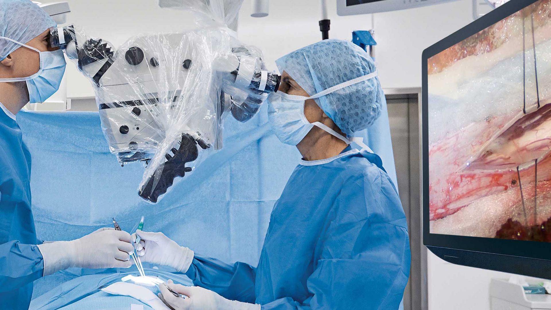
Leica Microsystems offers a range of surgical microscope solutions to support neurosurgeons in the most complex microsurgery applications.

Read More

Medical Device Manufacturing

Medical Device Manufacturing

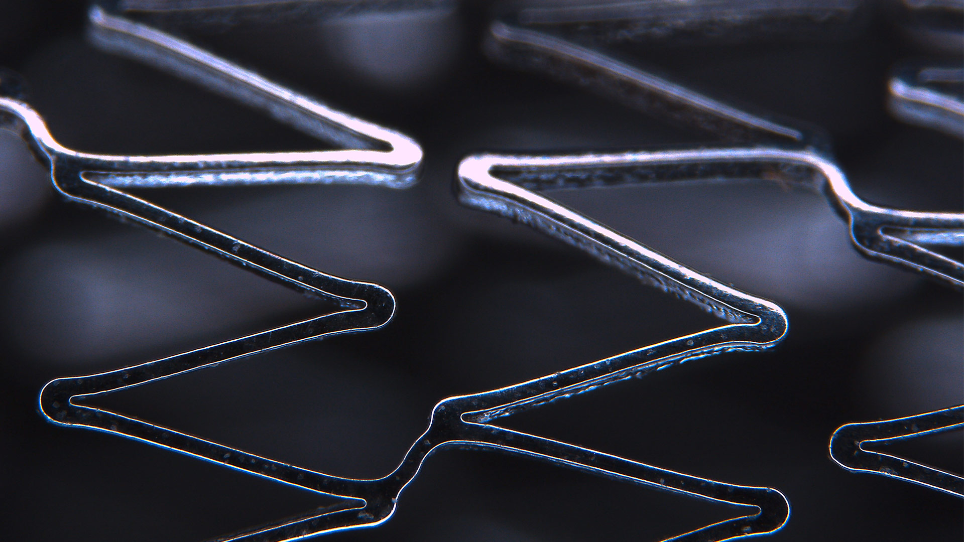
Medical device production or development requires Digital microscopes that deliver trustworthy image data ensuring that what you see is what you get. Identify, validate, and document quickly to help fulfill the compliance and safety requirements for your products and components.

Read More

Automotive and Transportation Sample

Automotive & Aerospace

Advances in e-mobility and navigation have made travel much easier. Fuel efficiency and safety are always improving. State-of-the-art communication and entertainment have dramatically changed our attitudes toward travel. Leica guides you to optimal imaging solutions for automotive and transportation, so you can stay ahead of the competition.

Read More

Mateo TL inside laminar flow hood user

Microscopy Solutions for Cell Culture

Get what you need and increase efficiency in your live cell imaging workflow with cell and tissue culture inverted microscopes from Leica Microsystems.

Read More

Neurology
Medical Device Manufacturing
Automotive and Transportation Sample
Mateo TL inside laminar flow hood user
Neurology
Medical Device Manufacturing
Automotive and Transportation Sample
Mateo TL inside laminar flow hood user
Neurology
Medical Device Manufacturing
Scroll to top