Overcoming Pediatric Brain Tumor Surgery Challenges
There are several challenges associated with achieving the safe and complete resection of brain tumors including ensuring adequate illumination and magnification, operating through safe surgical corridors to avoid neurological deficits, and precisely locating and determining the extent of brain tumors.
High resolution 4K surgical microscopes have significantly improved visualization while intra-operative guidance helps locate the tumor and guide tumor surgery to higher precision levels. This includes image-guided surgery, ultrasound-guided surgery and intra-operative MRI.
Improving Resection Precision with Fluorescence
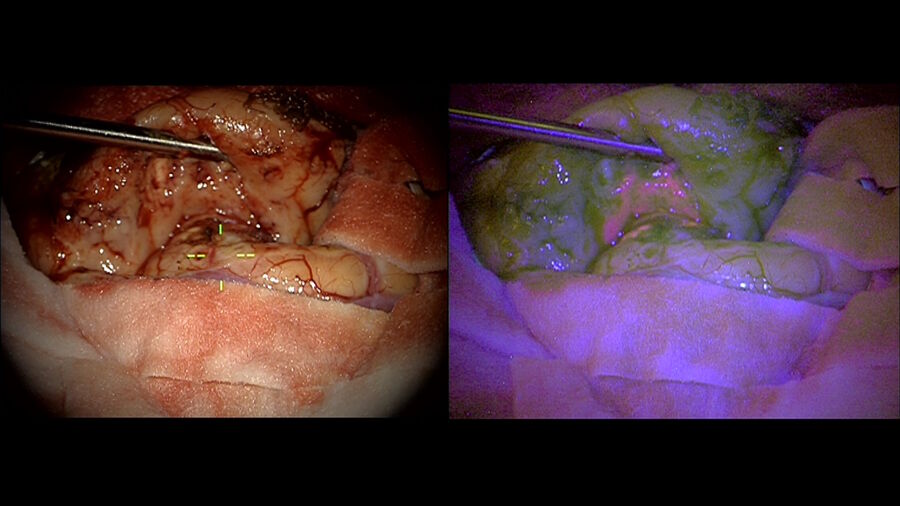
Fluorescence now offers a new level of precision. The FL400 fluorescence filter from Leica Microsystems used with 5-ALA during resection procedures, as experienced by Dr. Solanki, allows precise differentiation of tumor tissue from healthy brain tissue, at the touch of a button. Intense red fluorescence corresponds to solid tumor tissue and vague pink fluorescence to infiltrating tumor cells. Violet-blue light signals normal brain tissue.
In high-grade gliomas in adults, tumor fluorescence using 5-ALA reportedly improves the extent of resection compared to conventional white light surgery, whether or not guided by MRI-based navigation1. In pediatric brain tumors, Dr. Solanki recounts, high grade gliomas are also the most responsive to 5-ALA, and it is estimated that circa 90% of such tumors will show fluorescence.
5-ALA is administered orally around three hours before anesthesia. It is important to evaluate liver, renal and cardiac risks for patients, Dr. Solanki stresses.. Drug interactions should also be considered, for example with antiepileptic drugs which can reduce the presence of visible fluorescence in some tumors.
Use of 5-ALA in Pediatric Brain Tumors
Dr. Solanki explains that there is good evidence that 5-ALA fluorescence-guided surgery leads tohigher gross total resection (GTR) and progression free survival rate in high-grade gliomas in adults. Similar responses are noted in pediatric brain tumors. Studies on 5-ALA fluorescence-guided surgery in pediatric brain tumors found that 5-ALA-guided resection enabled the surgeon to identify the tumor more easily.
It was considered most helpful in cases of glioblastoma, anaplastic ependymoma and anaplastic astrocytoma. In contrast, the usefulness of fluorescence was found to be inconsistent in cases of PNETs, gangliogliomas, medulloblastomas and pilocytic astrocytomas.
The accumulation of fluorescent porphyrins seems to depend on WHO (World Health Organization) tumor grading. When 5-ALA-guided resections were considered helpful, the degree of resection was higher. The rate of adverse events related to 5-ALA was negligible.
It is critical to take precautions to avoid neurological deficits. 5-ALA-induced fluorescence of brain tissue does not provide information about the tissue’s underlying neurological function. Therefore, resection of fluorescing tissue should be weighed up carefully against the neurological function of fluorescing tissue.
Improving Visualization in 5-ALA-Based Fluorescence-Guided Surgery
Dr. Solanki also presents a point of view according to which using an advanced surgical microscope, such as the ARveo 8 or M530 OHX, equipped with blue light filters and 5-ALA fluorescence for enhanced intra-operative visualization with , has become an indispensable surgical technique setting a new standard of care for resection of high-grade gliomas in adults.
Dr. Solanki also presents a point of view according to which using an advanced surgical microscope, such as the ARveo 8 or M530 OHX, equipped with blue light filters and 5-ALA fluorescence for enhanced intra-operative visualization with , has become an indispensable surgical technique setting a new standard of care for resection of high-grade gliomas in adults.
Their role is also increasingly recognized in pediatric brain tumors. Pediatric neurosurgery and oncological units around the world, such as the Birmingham Women’s & Children’s Hospital, are increasingly using their added guidance for (more) precise resection of selective brain tumors.
Want to learn more? Register below to watch the full webinar presented by Dr. Guirish Solanki and see the different clinical cases he shared.
Note: Please note that off-label uses of products may be discussed. Please check with regulatory affairs for cleared indications for use in your region. The statements of the healthcare professionals included in this symposium reflect only their opinion and personal experience and not those of Leica Microsystems. They also do not necessarily reflect the opinion of any institution with whom they are affiliated.
Reference
- Optimization of high-grade glioma resection using 5-ALA fluorescence-guided surgery: literature review and practical recommendations from the neuro-oncology club of the French Society of Neurosurgery.


