
Introduction
Organoids, three-dimensional cell cultures that mimic the complexity of organs, have emerged as crucial models in cell biology and drug discovery. These structures provide a more physiologically relevant environment compared to traditional 2D cell cultures, allowing scientists to better understand organ development, disease mechanisms, and drug responses.
Organoids offer a unique advantage by replicating the cellular heterogeneity and architecture found in vivo. This is particularly valuable in studying diseases like cancer, where the microenvironment plays a crucial role. Additionally, organoids can be tailored to specific patient genotypes, facilitating personalized medicine research.
Challenges
Despite the advantages, observing and monitoring the growth and integrity of organoids pose challenges. Extracting organoids from culture systems without disrupting their structural integrity can be even more challenging. Maintaining the 3D architecture during harvesting and subsequent sample preparation for downstream analyses requires careful handling techniques.

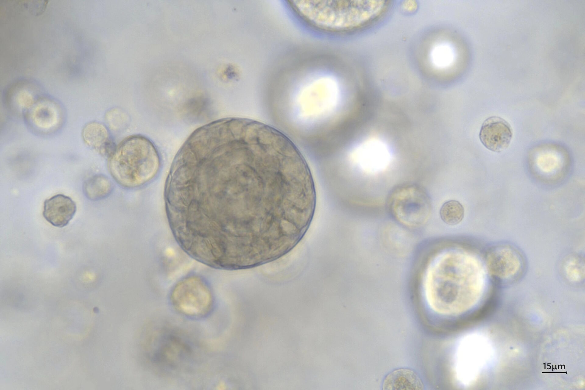
Courtesy of bioGenous, China.
Mateo TL Solution
Mateo TL digital microscope solution addresses these challenges by providing a platform for assessing imaging and documenting 3D cell cultures, especially organoids by allowing them to be studied within their culture systems. Moreover, the microscope allows the imaging of organoids within multi-well plates and microchips without the need for extraction or disruption. This capability addresses the challenges associated with maintaining the structural integrity of organoids during imaging, providing researchers with a non-invasive and efficient solution. With its compact design, Mateo TL fits inside a laminar flow hood enabling streamlined workflows under sterile conditions.

In this application note we highlight the following key benefits of using Mateo TL for the assessment of organoids:
1. Real-time monitoring
Mateo TL allows real-time monitoring of organoids directly within the multiwell plates and microchips. Researchers can observe dynamic changes, growth patterns, and cellular interactions without disturbing the 3D structure of the organoids over time, providing valuable insights into their development and response to experimental conditions.
2. Minimizing perturbation
The non-invasive nature of Mateo TL minimizes perturbation to the organoids. By eliminating the need for extraction or disruption, the system preserves the natural microenvironment and cellular interactions within the culture, ensuring that the observed data accurately reflects the in vivo-like conditions of the organoids. Using Mateo TL under the hood not only prevents contamination but also minimizes sample disturbance and streamlines workflow.
3. No compromise in image quality
Mateo TL's advanced optics and transmitted light imaging capabilities enable visualization of organoid morphology and cellular features within the multiwell plates or microchips. This level of precision is essential for capturing subtle changes and heterogeneity within the 3D structures without compromising image quality.

4. Multiple well inspection
With its versatile design, Mateo TL facilitates the inspection of multiple wells within the same experiment simultaneously. This feature streamlines the imaging process and allows researchers to analyze organoids across various conditions or treatments.
5. Workflow integration
Mateo TL is not just a standalone tool; it seamlessly integrates into a comprehensive imaging workflow. Researchers can leverage its capabilities as an initial observation step and subsequently transition to higher forms of imaging. The workflow progression might involve moving to advanced imaging systems like the THUNDER Imager, Mica, or STELLARIS confocal for high-resolution imaging, enabling detailed analysis of cellular structures and functions within organoids.

6. User-friendly interface
The user-friendly interface of Mateo TL simplifies the imaging process. Researchers can easily navigate through the multiwell plates, select regions of interest, and capture high-quality images, making it accessible for both novice and experienced users.
Conclusion
In conclusion, the Mateo TL digital microscope solution from Leica Microsystems offers effective means to help scientists overcome observational challenges in 3D cell cultures, particularly organoids. Its integration into a workflow solution empowers researchers to progress seamlessly from initial observations to higher-resolution imaging, facilitating a deeper understanding of organoid biology and advancing research in cell biology and drug discovery.

Courtesy of bioGenous, China.
 and phalloidin (magenta), imaged using Viventis SCAPE; scale bar 50μm. Courtesy of Marina Cuenca and Heleen Jungen (Dayton lab), EMBL Barcelona.")
 2D slices of a 1 mm diameter midbrain neural organoid stained with DAPI (blue, nuclear stain), β-tubulin (green, neuronal stain), and GFAP (red, astrocyte stain).")

