Leica Microsystems

Leica Microsystems

Leica Microsystems is a world leader in microscopes and scientific instruments. Founded as a family business in the nineteenth century, the company’s history was marked by unparalleled innovation on its way to becoming a global enterprise.

Its historically close cooperation with the scientific community is the key to Leica Microsystems’ tradition of innovation, which draws on users’ ideas and creates solutions tailored to their requirements. At the global level, Leica Microsystems is organized in three divisions, all of which are among the leaders in their respective fields: Life Science, Industry and Medical.

The company is represented in over 100 countries with 6 manufacturing facilities in 5 countries, sales and service organizations in 20 countries, and an international network of dealers. The company is headquartered in Wetzlar, Germany.
http://www.leica-microsystems.com/

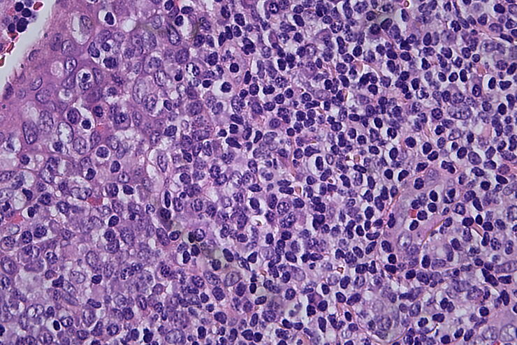
Masson-Goldner staining of a hedgehog brain slice.

How to Image Histological and Fluorescent Samples with One System

VIDEO ON DEMAND - How to image histological and fluorescent samples with one system. FluoSync, the new technology embedded into Mica enables the imaging of both histological staining and fluorescence…

Going Beyond Deconvolution

Widefield fluorescence microscopy is often used to visualize structures in life science specimens and obtain useful information. With the use of fluorescent proteins or dyes, discrete specimen…
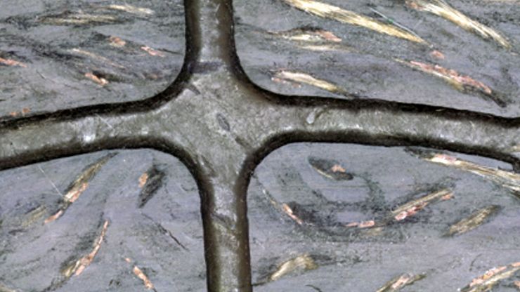
Inspection microscope image of a printed circuit board (PCB) taken with a ring light (RL) and near vertical illumination (NVI).

Microscope Illumination for Industrial Applications

Inspection microscope users can obtain information from this article which helps them choose the optimal microscope illumination or lighting system for inspection of parts or components.
Image of MDCK (Madin-Darby canine kidney) cells taken with phase contrast.

Phase Contrast and Microscopy

This article explains phase contrast, an optical microscopy technique, which reveals fine details of unstained, transparent specimens that are difficult to see with common brightfield illumination.

How to Radically Simply Workflows in Your Imaging Facility

VIDEO ON DEMAND - How to radically simplify imaging workflows and generate meaningful results with less time and effort using a highly automated microscope that unites widefield and confocal imaging.

Immersion Objectives

How an immersion objective, which has a liquid medium between it and the specimen being observed, helps increase the numerical aperture and microscope resolution is explained in this article.

Virtual Reality Showcase for STELLARIS Confocal Microscopy Platform

Take a seat directly next to our experts in front of the microscope, configure various multicolor experiments and see immediately the result. Be part of the highly interactive discovery tour of the…
Raw widefield and THUNDER image of Drosophila follicles. Image courtesy of M. Khoury and D. Bilder, University of California, Berkeley, USA.

Investigating Epithelial Morphogenesis and the Growth of Epithelia

This article shows how rapidly acquired THUNDER images of Drosophila follicles or egg chambers help distinguish apicobasal proteins when studying epithelial morphogenesis for cancer research.
Phase-contrast image of a MDCK-cell culture and its respective confluency measured by the Mateo TL microscope.

How to Determine Cell Confluency with a Digital Microscope

This article shows how to measure cell confluency in an easy and consistent way with Mateo TL, increasing confidence in downstream experiments.
Microscope image of cultured cells at the bottom of a dish.

How to do a Proper Cell Culture Quick Check

In order to successfully work with mammalian cell lines, they must be grown under controlled conditions and require their own specific growth medium. In addition, to guarantee consistency their growth…
Raw widefield and THUNDER image of GFP-tagged zebrafish fin. Courtesy of Jason Ear lab at Cal Poly Pomona, California, USA.

Diseases Linked to Scaffold Proteins and Signaling

This article shows how diseases related to scaffold proteins and protein signaling can be studied in zebrafish models efficiently with a THUNDER Imager.
Intensity distribution (arbitrary color coding) of an image of two points where the distance between them corresponds to the Rayleigh criterion.

Microscope Resolution: Concepts, Factors and Calculation

This article explains in simple terms microscope resolution concepts, like the Airy disc, Abbe diffraction limit, Rayleigh criterion, and full width half max (FWHM). It also discusses the history.
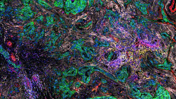
Pancreatic Ductal Adenocarcinoma with 11 cancer immunity and progression biomarkers shown –CD3, CD4, CD20, CD31, CD45, Collagen, HLA1, NaKATPase, PCK26, SMA1, and Vimentin.

Open Multiplexing: Transforming Tissue Research with Cell DIVE

Watch this webinar to discover how open multiplexing adapts to meet the needs of your multiplexing imaging study.
John Hamilton, Bchir, MB, FRCS, Consultant Ear, Nose & Throat Surgeon, Gloucester Royal Hospital. Dr. Hamilton during ontological surgery with his trainees.

Why 3D Microscope Visualization Enriches Surgical ENT Education

Read our new white paper to learn about the benefits of using a Leica surgical microscope with 3D visualization for surgical ENT education.
Extended depth of field reconstruction image of a whole human pancreas islet showing fluorescence signals from insulin (green), glucagon (red), an IL17 cytokine (magenta), and nuclei (blue).

Understanding Better the Onset of Diabetes

This article shows how Interleukin-17 (IL-17) proinflammatory cytokine proteins in human pancreatic islets can be studied efficiently with a THUNDER Imager.

Confocal Imaging of Immune Cells in Tissue Samples

In this webinar, you will discover how to perform 10-color acquisition using a confocal microscope. The challenges of imaged-based approaches to identify skin immune cells. A new pipeline to assess…

FluoSync - a Fast & Gentle Method for Unmixing Multicolour Images

In this white paper, we focus on a fast and reliable method for obtaining high-quality multiplex images in fluorescence microscopy. FluoSync combines an existing method for hybrid unmixing with…
Electric car or EV car charging in station. Eco-friendly alternative energy concept.

Alternative Fuels and Why Sustainable Solutions are Important

This free on-demand webinar is about the role of alternative fuel vehicles and why sustainable solutions are of increasing importance to the automotive industry.
Scroll to top