
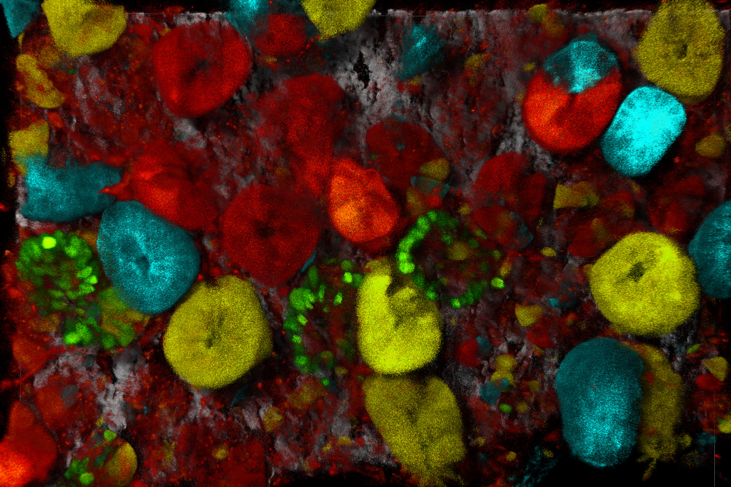
![[Translate to chinese:] Virally labeled neurons (red) and astrocytes (green) in a cortical spheroid derived from human induced pluripotent stem cells. THUNDER Model Organism Imagerwith a 2x 0.15 NA objective at 3.4x zoomwas used to produce this 425 μm Z-stack (26 positions), which is presented here as an Extended Depth of Field(EDoF)projection.](/fileadmin/_processed_/e/c/csm_THUNDER_Imager_Model-Org_Header-Gallery-Neuroscience_4652e132e6.jpg "[Translate to chinese:] Virally labeled neurons (red) and astrocytes (green) in a cortical spheroid derived from human induced pluripotent stem cells.")
Loading...
细胞生物学图片库
细胞生物学研究细胞的结构、功能和行为,包括细胞新陈代谢、细胞周期和细胞信号传导。荧光显微镜是细胞生物学家工具包中不可或缺的一部分。宽场显微镜和共聚焦显微镜被广泛用于观察细胞内复杂结构的细节。
Loading...

Developmental Biology Image Gallery
Developmental biology explores the development of complex organisms from the embryo to adulthood to understand in detail the origins of disease. This category of the gallery shows images about…