Loading...
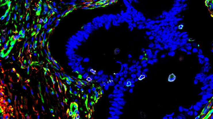
A Meta-cancer Analysis of the Tumor Spatial Microenvironment
Learn how clustering analysis of Cell DIVE datasets in Aivia can be used to understand tissue-specific and pan-cancer mechanisms of cancer progression
Loading...
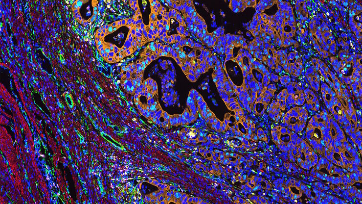
Mapping the Landscape of Colorectal Adenocarcinoma with Imaging and AI
Discover deep insights in colon adenocarcinoma and other immuno-oncology realms through the potent combination of multiplexed imaging of Cell DIVE and Aivia AI-based image analysis
Loading...
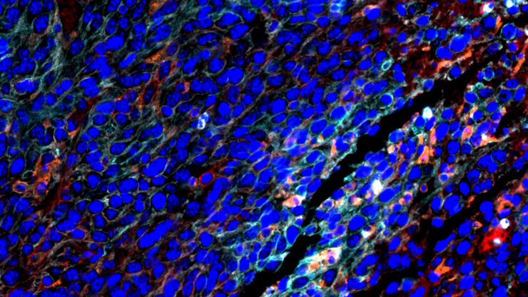
Spatial Architecture of Tumor and Immune Cells in Tumor Tissues
Dig deep into the spatial biology of cancer progression and mouse immune-oncology in this poster, and learn how tumor metabolism can effect immune cell function.
Loading...
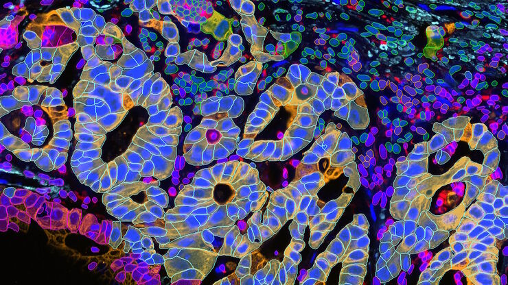
Transforming Multiplexed 2D Data into Spatial Insights Guided by AI
Aivia 13 handles large 2D images and enables researchers to obtain deep insights into microenvironment surrounding their phenotypes with millions of detected objects and automatic clustering up to 30…
Loading...
![[Translate to chinese:] Single cell datasets [Translate to chinese:] Single cell datasets](/fileadmin/_processed_/8/8/csm_Single_cell_datasets_SPARCS_441a58445d.jpg)
利用 SPARCS 探索亚细胞空间表型
功能日益强大的显微镜可提供信息丰富的各种细胞表型数据。如果与深度学习的最新进展相结合,这将成为在基因筛选中读出感兴趣的生物表型的理想技术。在本网络讲座中,您将了解到空间分辨 CRISPR 筛选 (SPARCS),这是一种利用自动化高速激光显微切割技术在人类基因组尺度上揭示各种亚细胞空间表型的平台。
Loading...
![[Translate to chinese:] THY1-EGFP labeled neurons in mouse brain processed using the PEGASOS 2 tissue clearing method. Neurons were traced using Aivia’s 3D Neuron Analysis – FL recipe. Image credit: Hu Zhao, Chinese Institute for Brain Research. [Translate to chinese:] THY1-EGFP labeled neurons in mouse brain processed using the PEGASOS 2 tissue clearing method, imaged on a Leica confocal microscope. Neurons were traced using Aivia’s 3D Neuron Analysis – FL recipe. Image credit: Hu Zhao, Chinese Institute for Brain Research.](/fileadmin/_processed_/f/1/csm_Neurons_in_mouse_brain_with_Aivia_3D_Neuron_Analysis_9aa755e1c5.jpg)
借助人工智能,揭示复杂而密集的神经元图像中的洞察
神经元的3D形态学分析通常需要使用不同的成像模式,捕捉多种类型的神经元,并在各种密度下相连的传统Leica SP8显微镜采集多达解神经元的形态,这对许多研究人员来说仍然是一个耗时的挑战。
Loading...
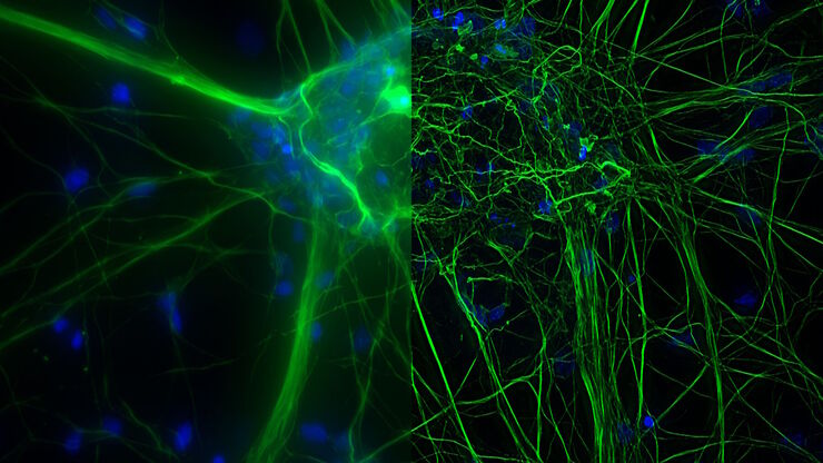
What are the Challenges in Neuroscience Microscopy?
eBook outlining the visualization of the nervous system using different types of microscopy techniques and methods to address questions in neuroscience.
Loading...
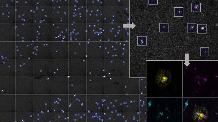
人工智能显微成像能够高效检测稀有事件
对稀有事件进行定位和选择性成像是许多生物样本研究过程的关键。然而,由于时间限制和高度的复杂性,有些实验无法做到,从而限制了获得新发现的前景。通过基于人工智能的显微成像检测稀有事件,这种工作流程将智能样本导航、图像采集工具和人工智能驱动的图像分析等不同功能融合起来共同协作,能够克服上述局限性。
Loading...
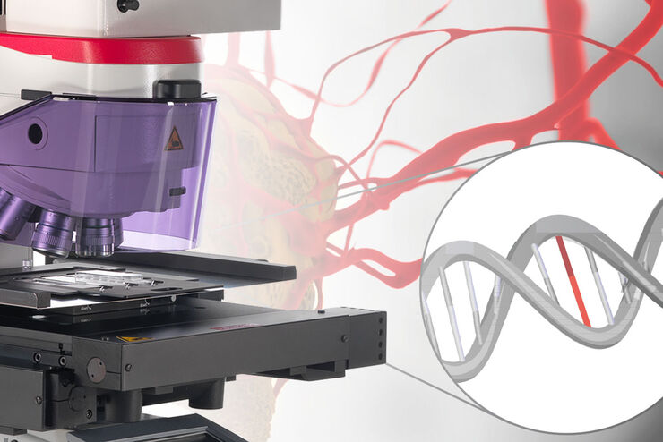
组织中的精密空间蛋白质组学信息
尽管可使用基于成像和质谱的方法进行空间蛋白质组学研究,但是图像与单细胞分辨率蛋白丰度测量值的关联仍然是个巨大的挑战。最近引入的一种方法,深层视觉蛋白质组学(DVP),将细胞表型的人工智能图像分析与自动化的单细胞或单核激光显微切割及超高灵敏度的质谱分析结合在了一起。DVP在保留空间背景的同时,将蛋白丰度与复杂的细胞或亚细胞表型关联在一起。
