Loading...
![[Translate to chinese:] Microscope equipped with a K7 color CMOS camera for life-science and industry imaging applications.](/fileadmin/_processed_/9/e/csm_Microscope_equipped_with_a_K7_color_CMOS_d4485ce44f.jpg "[Translate to chinese:] Microscope equipped with a K7 color CMOS camera for life-science and industry imaging applications.")
数码显微镜摄像头和图像分析的基础技术术语定义
现今绝大多数显微镜都配置了摄像头。摄像头的特征通常决定了所采集到的图像是否能够揭示出研究人员希望观察到的现象。但深入到摄像头术语时,技术术语十分繁杂。我们汇总整理了最为重要的术语及其简明释意以便提供方向。这些术语按字母顺序排列。
Loading...

Epi-Illumination Fluorescence and Reflection-Contrast Microscopy
This article discusses the development of epi-illumination and reflection contrast for fluorescence microscopy concerning life-science applications. Much was done by the Ploem research group…
Loading...
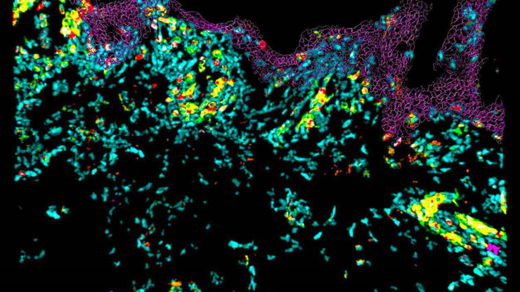
![[Translate to chinese:] C. elegans adult hermaphrodite gonades acquired using THUNDER Imager. Staining: blue - DAPI (nucleus), green - SP56 (sperm), red - RME-2 (oocyte), magenta - PGL-1 (RNA + protein granules). Image courtesy of Prof. Dr. Christian Eckmann, Martin Luther University, Halle, Germany.](/fileadmin/_processed_/7/c/csm_C_elegans_adult_hermaphrodite_gonades_6194ed03fc.jpg "[Translate to chinese:] C. elegans adult hermaphrodite gonades acquired using THUNDER Imager. Image courtesy of Prof. Dr. Christian Eckmann, Martin Luther University, Halle, Germany.")
生命科学研究: 哪种显微镜相机适合您?
相机是显微镜系统的重要组成部分,对系统的性能有重大影响。在选择相机时,重要的是不仅要看技术规格,还要考虑您的样品、技术、对比方法以及您希望获得的数据类型。
Loading...
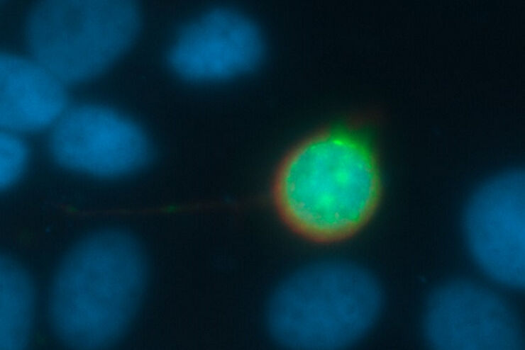
![[Translate to chinese:] Branched organoid growing in collagen where the Nuclei are labeled blue. To detect the mechanosignaling process, the YAP1 is labeled green.](/fileadmin/_processed_/a/e/csm_Branched_organoid_growing_in_collagen_083254eb44.jpg "[Translate to chinese:] Branched organoid growing in collagen where the Nuclei are labeled blue. To detect the mechanosignaling process, the YAP1 is labeled green.")
检查癌症类器官的发展进程
德国慕尼黑工业大学的Andreas Bausch实验室研究细胞和生物体中不同结构和功能形成的细胞和生物物理机制。他的团队设计了新的策略、方法和分析工具,以量化微米和纳米等级的发展机制和动态过程。关键研究领域包括干细胞和类器官,从乳腺类器官到胰腺癌类器官,以更好地了解疾病模型。
Loading...
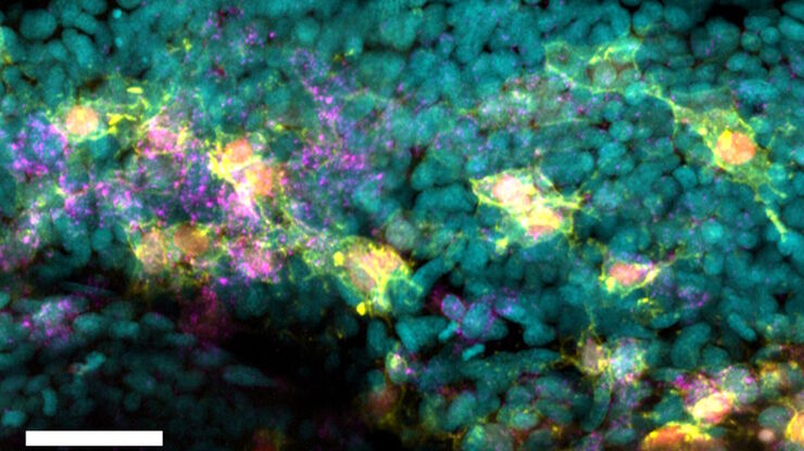
![[Translate to chinese:] Raw widefield and THUNDER image of calcium transients in Drosophila embryos. Courtesy A. Carreira-Rosario, Clandinin laboratory, California, USA.](/fileadmin/_processed_/7/6/csm_Calcium_transients_in_Drosophila_embryos_teaser_fcd31bd78f.jpg "[Translate to chinese:] Raw widefield and THUNDER image of calcium transients in Drosophila embryos. Courtesy A. Carreira-Rosario, Clandinin laboratory, California, USA.")
Central Nervous System (CNS) Development and Activity in Organisms
This article shows how studying central nervous system (CNS) development in Drosophila-melanogaster embryos expressing a GCaMP calcium indicator in the neurons can be improved with a THUNDER Imager.
Loading...

How to Image Histological and Fluorescent Samples with One System
VIDEO ON DEMAND - How to image histological and fluorescent samples with one system. FluoSync, the new technology embedded into Mica enables the imaging of both histological staining and fluorescence…
Loading...
Going Beyond Deconvolution
Widefield fluorescence microscopy is often used to visualize structures in life science specimens and obtain useful information. With the use of fluorescent proteins or dyes, discrete specimen…
Loading...
How to Radically Simplify Workflows in Your Imaging Facility
VIDEO ON DEMAND - How to radically simplify imaging workflows and generate meaningful results with less time and effort using a highly automated microscope that unites widefield and confocal imaging.