How to Radically Simplify Workflows in Your Imaging Facility

MicaCam episode 9 - Video On Demand

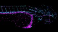
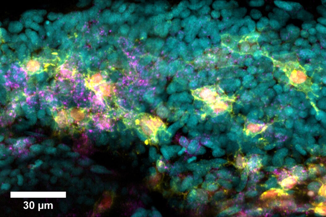
 Fixed_zebrafish_embryos_in_Agarose_PBS.jpg

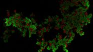
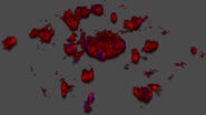
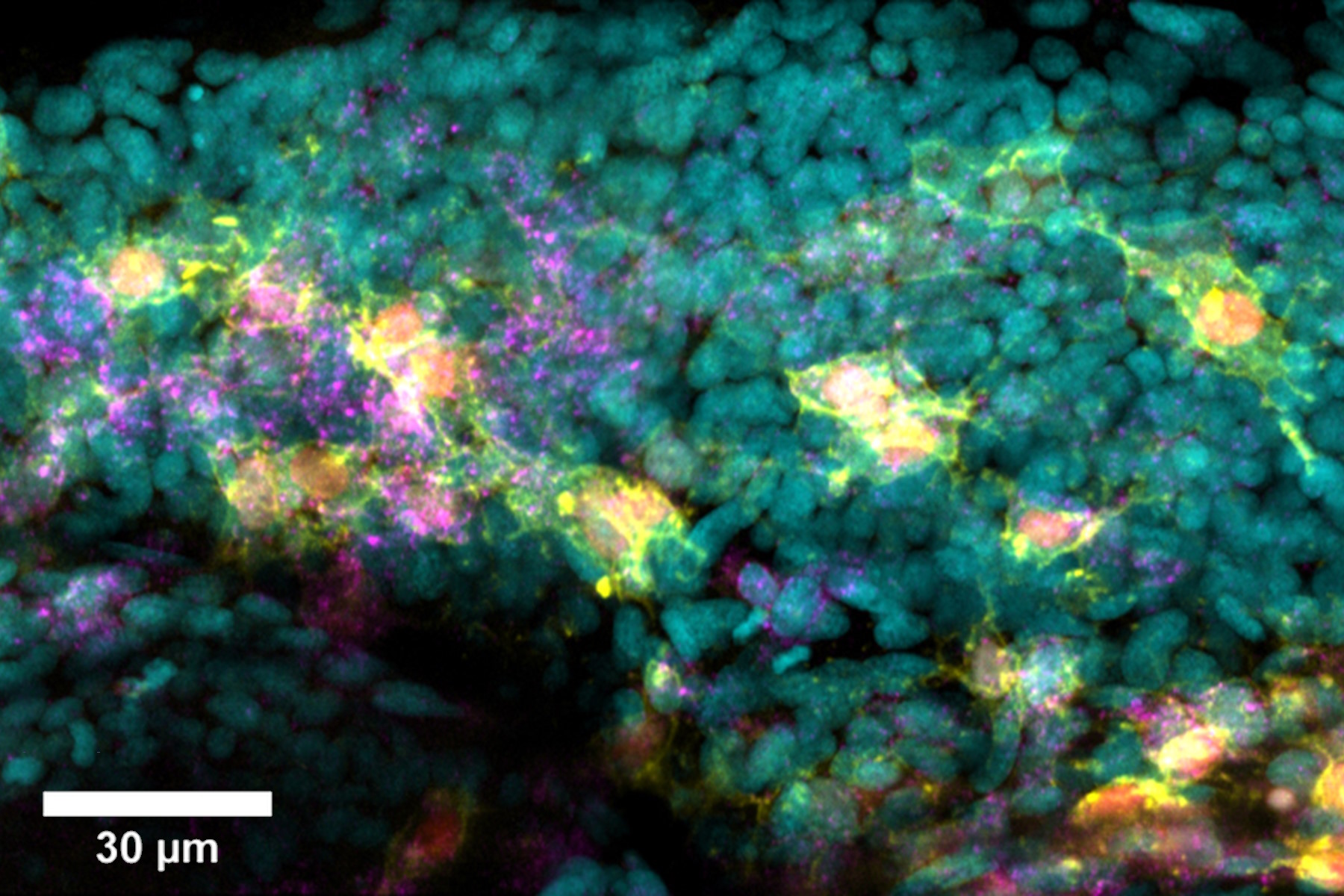
In this episode of MicaCam, our special guest Dr Christopher Thrasivoulou from the University College London (UCL) will discuss the benefits of utilizing Mica from an Imaging Facility perspective. He will talk about automated and streamlined imaging workflows for complex biological systems. These help scientists to generate meaningful quantitative results with less time and effort. He will present an example of such a workflow by performing multi-color imaging of fluorescently labelled, fixed zebrafish embryos.

To watch the full MicaCam experiment, simply fill in a few details in the form below to get your video on demand, and access even more exclusive experiments in the MicaCam library.

Watch now!

WHAT TO EXPECT IN THE WEBINAR

Key Learnings

  • How to set up a streamlined imaging workflow for complex biological systems such as zebrafish embryos
  • How to capture multiple labels of different structures in a single acquisition either using widefield or confocal imaging
  • How to save both imaging and training time using Intelligent Automation

Speakers

Dr. Lynne Turnbull

Principal Scientist - Leica Labs @ EMBL Imaging Center

Lynne is a Principal Scientist at Leica Microsystems. She received her PhD in Sydney Australia in cardiac biophysics and undertook postdoctoral training in San Francisco and Melbourne. Lynne’s research interests shifted to bacterial biofilms and motility, and she used different types of imaging to explore and understand how bacteria build communities and move through their environment. Upon moving to the University of Technology Sydney, Lynne established and managed the Microbial Imaging Facility. Lynne joined GE in 2016 to provide application support throughout Asia for super resolution microscopy. Since 2021 Lynne has been with Leica Microsystems based in the labs at the EMBL Imaging Center in Heidelberg.

Dr. Christopher Thrasivoulou

PhD. FRMS. Director of UCL’s Division of Biosciences Imaging Facility

Chris obtained his PhD from University College London in 1998 and is currently Director of UCL’s Division of Biosciences Imaging Facility. This large state-of-the -art imaging unit serves over 900 researchers from a very diverse range of science disciplines across the whole of UCL and number of external UK universities. The unit has 10 confocal microscopes, including super-resolution confocals, 2-Photon, SIM/PALM/STORM/TIRF, Lighsheet, High content imaging, Slide scanner, Raman and X-Ray CT.  Chris’s research interest began in the field of neuroanatomy, studying the effects of aging and caloric restriction in the central and peripheral nervous system at the Royal Free Hospital School of Medicine. Chris moved to UCL in 2002, his research being focused on studying the role of connexins in wound healing and cancer cell biology and the role Wnt/calcium signalling in cancer aetiology.
REGISTER TO WATCH THE EXPERIMENTS NOW

By clicking on the SUBMIT button, I confirm that I have reviewed and agree with Leica Microsystems GmbH Terms of Use and their Privacy Policy. I also understand my privacy choices as they pertain to my personal data, as detailed in the aforementioned Privacy Policy under ‘’Your Privacy Choices’’.

Related Articles

Background image
Scroll to top