Contact Us

How to Image Histological and Fluorescent Samples with One System

MicaCam Episode 10 - Video on demand

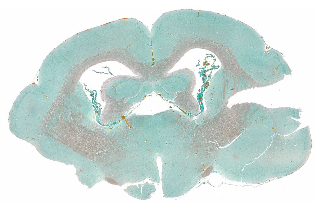
Masson-Goldner staining of a hedgehog brain slice. Masson-Goldner_staining_of_hedgehog_brain_slice.jpg

In this episode of MicaCam, our hosts Lynne Turnbull and Patric Pelzer will take you on a journey through the history of staining biological samples. They will explain why you typically have to make a choice to either use a system for histology or for fluorescent samples and how you can overcome this with the new imaging technique – FluoSync.

MicaCam is where life science researchers come together live to chat, interact, and make discoveries together. Share your questions and engage during the livestream. 
Join us, and your life science research community, for short demonstrations of how Mica radically simplifies your workflows.

WATCH NOW!

What to expect in the webinar

Key Learnings

  • What is the difference between colorimetric/RGB imaging and fluorescence imaging?
  • How to image both histological staining and fluorescence without compromise thanks to the FluoSync technology
Register to watch the experiments now

By clicking on the SUBMIT button, I confirm that I have reviewed and agree with Leica Microsystems GmbH Terms of Use and their Privacy Policy. I also understand my privacy choices as they pertain to my personal data, as detailed in the aforementioned Privacy Policy under ‘’Your Privacy Choices’’.

Related Articles

Background image
Scroll to top