Advancing 3D culture imaging
Unleashing the power of microscopy
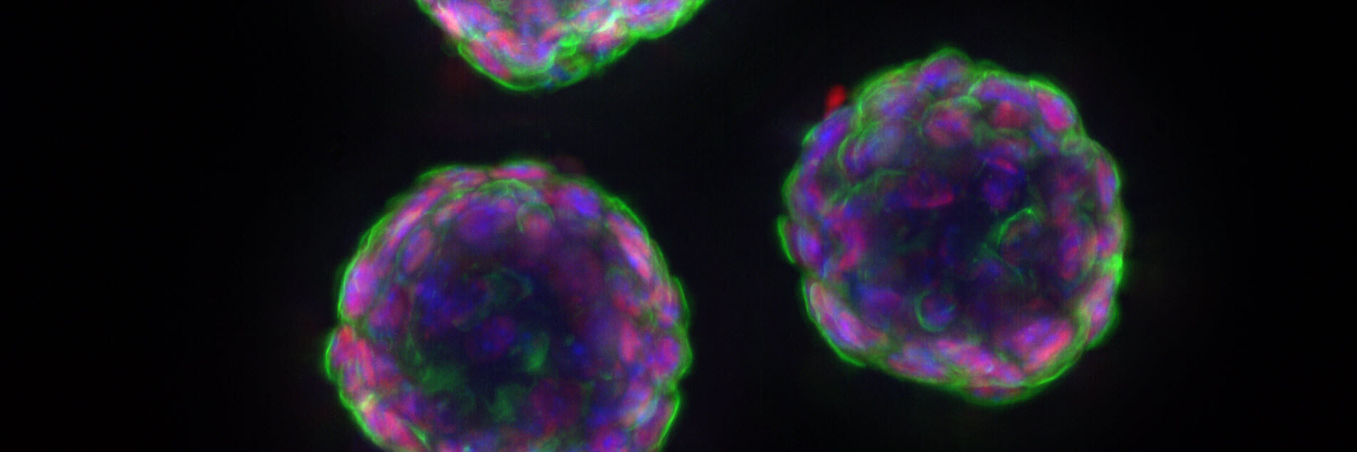
Organoids, spheroids, and other 3D culture models have become indispensable tools for life science research, allowing researchers to better study physiological processes such as development, homeostasis, regeneration and disease. However, imaging these types of models presents unique challenges and practical imaging solutions are crucial if deeper insights are to be gained.
This eBook examines some of the challenges encountered when imaging 3D culture models, and sheds light on innovative microscopy solutions that can empower scientists to make new advances in areas such as regenerative medicine, drug discovery, and disease research.
We focus on several case studies including:
- Examining ‘Brains-In-A-Dish’ from induced pluripotent stem cells (iPSCs)
- Observing 3D cell cultures during development
- Developing heart pacemaker cells from cardiac spheroids
Learn more about:
- Key considerations for imaging organoids and spheroids
- Solutions for examining dynamic processes in organoids and spheroids in real-time
- Going deeper into 3D with correlative light and electron microscopy (CLEM)