
Loading...
将动态活细胞数据融入超微结构
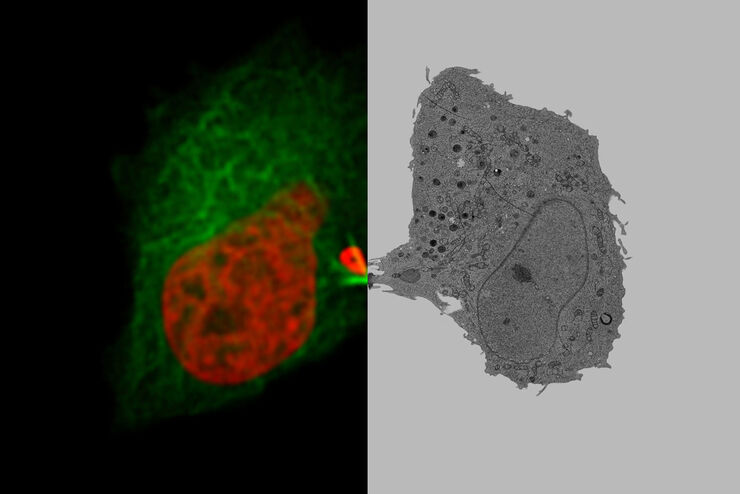
采用徕卡Nano的工作流程,可以避免过去如海底捞针似的寻找。利用光电关联显微技术,在适当的时间直接鉴别出正确的细胞,并将动态的活细胞数据融入其超微结构中。
Loading...
Advancing Cell Biology with Cryo-Correlative Microscopy
Correlative light and electron microscopy (CLEM) advances biological discoveries by merging different microscopes and imaging modalities to study systems in 4D. Combining fluorescence microscopy with…
Loading...

Development of Fluorescence Lifetime Imaging Microscopy (FLIM) and its Relevance for Functional Imaging
Prof. Ammasi Periasamy, Director, Keck Center for Cellular Imaging, University of Virginia, was interviewed by Dr. Giulia Ossato, Product Manager functional imaging, during Leica Microsystems Meets…
Loading...
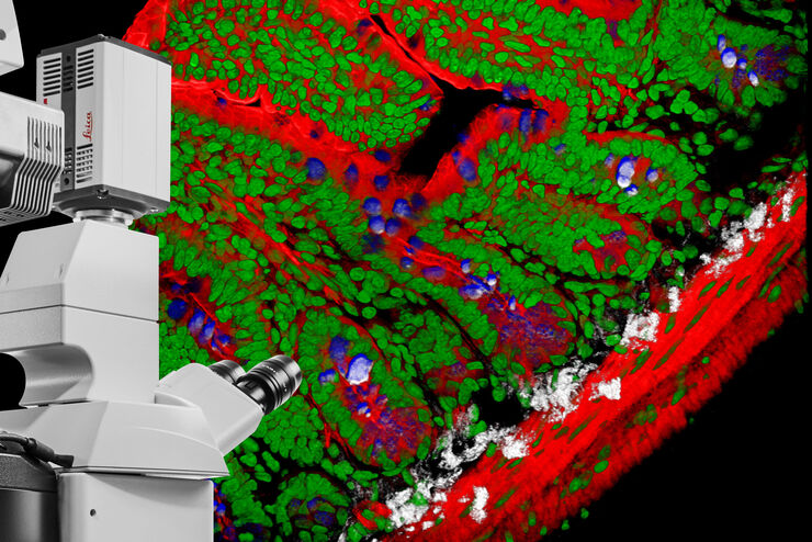
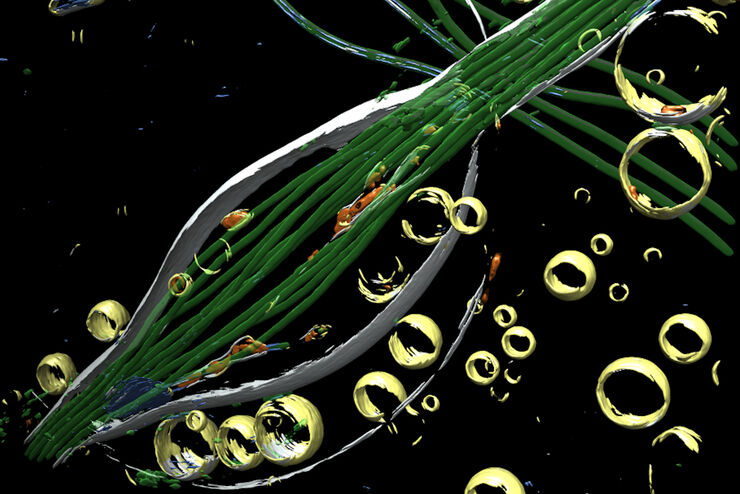
DIVE Multiphoton Microscope Image Gallery
Today’s life science research focusses on complex biological processes, such as the causes of cancer and other human diseases. A deep look into tissues and living specimens is vital to understanding…
Loading...
![[Translate to chinese:] Mammalian cell culure. Phase contrast and fluorescence image.](/fileadmin/_processed_/3/0/csm_Mammalian_cells_culture_teaser_7ea370b52b.jpg "[Translate to chinese:] Mammalian cell culure. Phase contrast and fluorescence image.")
哺乳动物细胞培养的介绍
哺乳动物细胞培养是生命科学的基本支柱之一。如果不具备在实验室中培养细胞的能力,那么细胞生物学、免疫学、肿瘤研究等学科很难实现快速发展。本文概述了哺乳动物细胞培养系统,可以根据其形态、细胞类型和组织对其进行分类。此外,还介绍了适宜的细胞生长条件以及需要使用何种显微镜来观察细胞。
Loading...

About the Most Important Considerations When Imaging Deep Into Mouse Tissue
When operating a confocal microscope, or when discussing features and parameters of such a device, we inescapably mention the pinhole and its diameter. This short introductory document is meant to…
Loading...

Gene Editing with CRISPR/Cas9 - Breakthrough in Genome Engineering
The CRISPR/Cas9 system is one of several different bacterial systems for defense against viral attacks. It consists of two main components. One is a small piece of RNA which binds to the viral target…
Loading...
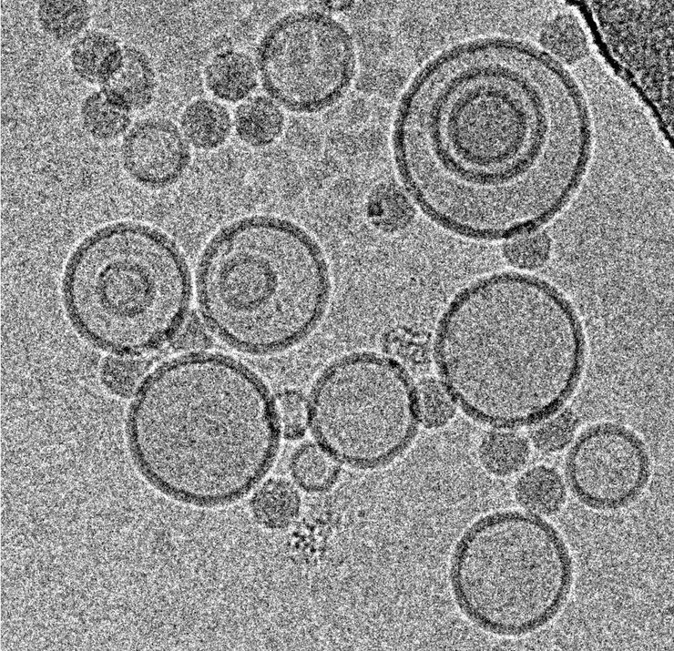
冷冻透射电子显微镜的投入式冷冻技术:应用
低温下观察完全含水、未染色样本的透射电子显微镜(cryo TEM)是结构生物学、细胞生物学、药理学和其他科学分支的通用工具。通过将标本放入冷冻剂中进行超快速冷冻(投入式冻结)是一种常用的方法,用于制备在透射电镜观察的各种标本。本文是对投入式冷冻的补充,介绍了在不同领域使用投入式冷冻标本的三种冷冻TEM应用。
Loading...
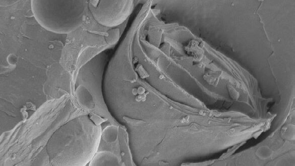
冷冻断裂与冷冻蚀刻基础介绍
冷冻断裂是一种将冰冻样本劈裂以露出其内部结构的技术。冷冻蚀刻是指让样本表面的冰在真空中升华,以便露出原本无法观察到的断裂面细节。金属/碳复合镀膜能够实现样本在SEM(块面)或TEM(复型)中的成像,主要用于研究如细胞器、细胞膜,细胞层和乳胶。这项技术传统上用于生物学应用,但现在逐渐在物理学和材料科学中展现出重要意义。近年来,研究人员通过冷冻断裂电子显微镜,尤其是冷冻复型免疫标记(FRIL),对膜蛋…