
Loading...
![[Translate to chinese:] Raw widefield and THUNDER image of transversal mouse adult fiber lens section. Courtesy N. Houssin, Plagemen lab, Ohio State University, Columbus, USA.](/fileadmin/_processed_/3/0/csm_Transversal_mouse_adult_fiber_lens_section_Widefield_THUNDER_teaser_6f95700ba0.jpg "[Translate to chinese:] Raw widefield and THUNDER image of transversal mouse adult fiber lens section. Courtesy N. Houssin, Plagemen lab, Ohio State University, Columbus, USA.")
眼部出生缺陷研究
本文讨论如何使用快速采集的宽场显微镜图像研究晶状体的形成和眼部出生缺陷。将小鼠晶状体用作模型,以研究眼睛细胞形态发生及细胞过程如何中断并导致发生缺陷。通过无模糊或离焦模糊的图像揭示了小鼠晶状体玻片内部深处的细节。这些图像是使用THUNDER Imager 3D Assay采集的。
Loading...
![[Translate to chinese:] Images of smooth muscle cells during wound healing. Courtesy L.S. Shankman, Ph.D., University of Virginia.](/fileadmin/_processed_/4/3/csm_Smooth_muscle_cells_during__26769ad3cd.jpg "[Translate to chinese:] Images of smooth muscle cells during wound healing. Courtesy L.S. Shankman, Ph.D., University of Virginia.")
平滑肌细胞划痕愈合研究
本文主要讨论如何使用专门配置的徕卡倒置显微镜和台式细胞培养箱轻松、可靠地研究多孔板中培养的平滑肌细胞(SMC)的划痕愈合情况。血管受损后影响SMC增殖和迁移的信号转导情况在医学研究中有重要意义。由于SMC遍布全身,所以对其迁移情况的研究也有助于癌症和损伤的治疗。
Loading...

How AR Helps in the Surgical Treatment of Moyamoya Disease
Moyamoya disease is a rare chronic occlusive cerebrovascular disorder characterized by progressive stenosis in the terminal portion of the internal carotid artery and an abnormal vascular network at…
Loading...
![[Translate to chinese:] Clinical case: radial forearm free flap preparation with prelaminated urethra and phalloplasty anastomosis](/fileadmin/_processed_/b/e/csm_Urethra_and_phalloplasty_anastomosis_0b94d56aa8.jpg "[Translate to chinese:] Urethra and phalloplasty anastomosis")
增强现实荧光技术在桡侧前臂皮瓣游离阴茎成形术中的应用
这个手术中,首席显微外科医生教授及其团队进行了桡侧前臂游离皮瓣阴茎成形术,并使用ICG荧光成像来显示整个皮瓣中从吻合口到阴茎远端的血流。教授展示了增强现实荧光技术除了常见的临床症状外,还能提供检查血流的状态。
Loading...
 expressed in the sensory neurons.")
感觉神经元的高对比度快速三维成像
本文讨论了相比传统的宽场显微镜,使用large volume computational clearing(LVCC)技术的THUNDER组织成像系统如何获取背根神经节(DRG)组织高对比度的快速三维成像图,获得感觉神经元更为清晰的解析图像。神经科学研究的一项主要领域集中在感觉神经元对触觉和痛觉的影响方面。深入理解这种现象对于神经系统疾病和疗法的发展具有重要意义。
Loading...
 or Minor’s syndrome")
Minor’s Syndrome Surgical Intervention by Prof. Vincent Darrouzet
Minor’s disease, also called Superior Semicircular Canal Dehiscence (SSCD) or Minor’s syndrome, is a rare disorder of the inner ear that affects hearing and balance. The disease is characterized by…
![[Translate to chinese:] Raw widefield (left) and THUNDER (right) image of Ewing Sarcoma cells (SK-ES-1).](/fileadmin/academy/2022/Visualizing_the_Mitotic_Spindle_in_Cancer_Cells/Tubulin_mitotic_spindle_teaser.jpg "[Translate to chinese:] Raw widefield (left) and THUNDER (right) image of Ewing Sarcoma cells (SK-ES-1)")
Loading...
Formulated Product Characterization with SRS Microscopy
Creams, pastes, gels, emulsions, and tablets are ubiquitous across a wide range of manufacturing sectors from pharmaceuticals and consumer health products to agrochemicals and paint. To improve…
Loading...
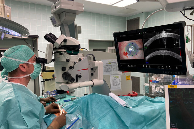
Proveo 8 with intraoperative OCT – a User Evaluation in an University Setting
Optical coherence tomography (OCT) makes structures in the eye visible that lie beneath the surface. When OCT is used intraoperatively, surgeons gain insight into possible pathological changes in the…