
Loading...

Advances in Oncological Reconstructive Surgery
Decision making and patient care in oncological reconstructive surgery have considerably evolved in recent years. New surgical assistance technologies are helping surgeons push the boundaries of what…
Loading...

精确分析宽视野荧光图像
利用荧光显微镜的特异性,即便是使用厚样品和大尺寸样品,研究人员也能够快速轻松地准确观察和分析生物学过程和结构。然而,离焦荧光会提高背景荧光,降低对比度,影响图像的精确分割。THUNDER 与Aivia 的组合可以有效解决这一问题。前者可以消除图像模糊,后者会使用人工智能技术自动分析宽视野图像,提高操作速度和精确性。下面,我们来详细了解下这一协作方法。
Loading...

Imaging of Anti-Cancer Drug Uptake in Spheroids using DLS
Spheroid 3D cell culture models mimic the physiology and functions of living tissues making them a useful tool to study tumor morphology and screen anti-cancer drugs. The drug AZD2014 is a recognized…
Loading...

目视检查面临的主要挑战
本文讨论使用显微镜进行目视检查和返工时遇到的挑战。使用正确类型的显微镜和光学设置对于优化工作流程和增加产量至关重要。使用显微镜进行目视检查和返工时可能遇到的挑战包括确定适当的放大倍率和照明以及有足够大的工作距离。然而,其他关键因素与工作流程优化、有效报告结果和用户培训以及检查过程中的用户舒适度有关。Leica数码和体视显微镜能够提供一系列完整的解决方案,帮助您克服这些挑战,提供更有效的检查和返工。
Loading...
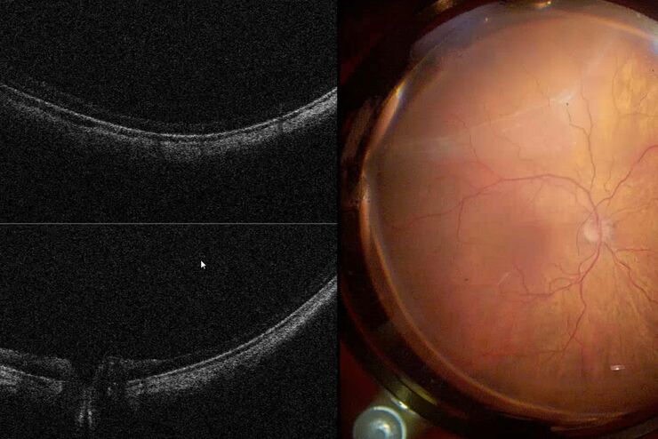
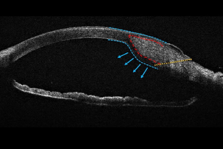
术中光学相干断层扫描(OCT)在视网膜手术中的应用
在过去的一年,巴西某视网膜诊所的手术团队采用了显微镜集成式术中OCT。在他们发布的白皮书《术中光学相干断层扫描:视网膜手术的临床经验》中,深入分析了使用OCT的复杂视网膜手术病例,其中OCT额外提供了有用信息,某些情况下这些信息对外科医生做出决策至关重要。
Loading...
![[Translate to chinese:] HeLa Kyoto cells (HKF1, H2B-mCherry, alpha Tubulin, mEGFP). Left image: Maximum projection of a z-stack prior to ICC and LVCC. Right image: Maximum projection of a mosaic z-stack after ICC and LVCC.](/fileadmin/_processed_/6/a/csm_How_to_improve_live_cell_imaging_with_Leica_Nano_Workflow_teaser_04b9da73e4.jpg "[Translate to chinese:] HeLa Kyoto cells")
如何使用Coral Life(活细胞光电联用)改进活细胞成像
对于活细胞 CLEM 应用而言,光学显微镜成像是在正确的时间以正确的状态识别正确细胞的关键步骤。在本文中,徕卡专家就使用宽场系统的优势以及使用蓝宝石作为细胞培养基底时需要克服的障碍分享了他们的见解。
Loading...
![[Translate to chinese:] The EM ICE Nano loading area](/fileadmin/_processed_/3/c/csm_Leica_EM_ICE_Samp-Link-Chamber_799472ccbb.jpg "[Translate to chinese:] The EM ICE Nano loading area")
如何让样品保持在生理状态
Coral Life工作流将动态数据与最佳的样品固定方式(高压冷冻)相结合。然而,如果您的细胞因为温度下降,或缺氧气、二氧化碳或营养物质缺乏而受到损伤,那么再好的样品保存也没有意义。这些因素将影响一系列的生物过程,甚至破坏原超微结构基础,影响您的分析。
Loading...
Towards Advanced Use of Intraoperative OCT in Cataract Surgery
In this White Paper, Dr. Rachid Tahiri shares his personal experience with the Leica EnFocus intraoperative OCT, the valuable features supporting smooth surgery and how it allows him to minimize…
![[Translate to chinese:] Virally labeled neurons (red) and astrocytes (green) in a cortical spheroid derived from human induced pluripotent stem cells. THUNDER Model Organism Imager with a 2x 0.15 NA objective at 3.4x zoom was used to produce this 425 µm Z-stack (26 positions), which is presented here as an Extended Depth of Field (EDoF) projection. Images courtesy of Dr. Fikri Birey from the Dr. Sergiu Pasca laboratory at Stanford University, 3165 Porter Dr., Palo Alto, CA](/fileadmin/_processed_/2/0/csm_Neural-sphere_model-org_LVC_61ecf44e40.jpg "[Translate to chinese:] Virally labeled neurons (red) and astrocytes (green) in a cortical spheroid derived from human induced pluripotent stem cells.")