
Loading...

术中OCT引导的青光眼支架修复手术
青光眼是导致全球不可逆失明的主要原因之一。小梁网切除术和导管分流引流术等历史悠久的手术技术会带来巨大的短期风险和潜在并发症。近年来,随着微创青光眼手术(MIGS)的出现,手术方法有了长足的发展,其特点是对组织的破坏最小、内路粘小管植入、手术时间短、器械简单、术后恢复快。
Loading...
![[Translate to chinese:] Dr. Ozana Moraru shares two primary open-angle glaucoma cases in which trabeculectomy bleb needling was performed using the Leica M844 microscope with EnFocus intraoperative OCT. Image courtesy of Dr. Ozana Moraru.](/fileadmin/_processed_/1/0/csm_Trabeculectomy_bleb_needling_performed_with_Leica_M844_microscope_with_EnFocus_intraoperative_OCT_81e3cbbf3e.jpg "[Translate to chinese:] Dr. Ozana Moraru shares two primary open-angle glaucoma cases in which trabeculectomy bleb needling was performed using the Leica M844 microscope with EnFocus intraoperative OCT. Image courtesy of Dr. Ozana Moraru.")
术中光学显像如何帮助青光眼手术获得更多洞察力
青光眼是导致全球失明的主要原因之一。青光眼手术可以延缓疾病的发展。在青光眼手术过程中,术中光学相干断层扫描(OCT)的使用为眼科外科医生提供了更佳的可视化效果,让他们更深入地了解表面下组织对手术操作的反应。 莫拉鲁博士通过两个原发性开角型青光眼(POAG)的临床病例强调了它的价值。
Loading...
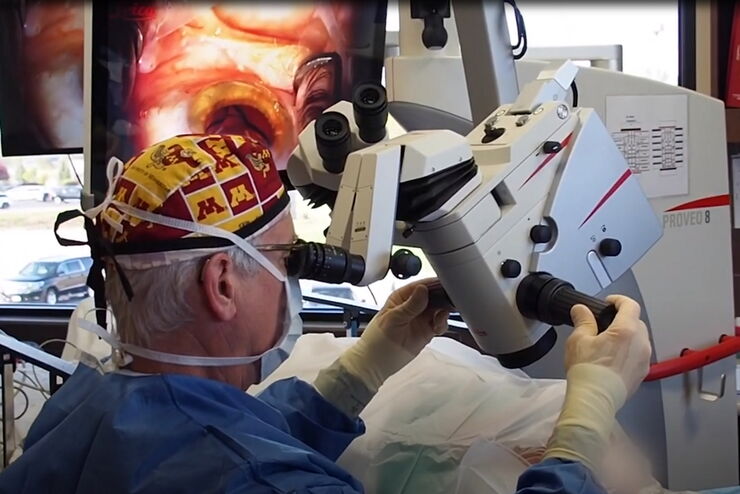
Improved Visualization of the Anterior Chamber Angle with Proveo 8
In this interview, Michael G. Richie, MD offers his expert opinion on the Proveo 8 from Leica Microsystems as a means for outstanding visualization of the anterior chamber during MIGS. He describes…
Loading...
Overcoming Ophthalmologic Surgery Challenges
Ophthalmology surgical procedures involving both the anterior and posterior segment can be particularly challenging. Good visualization is required to operate with precision and confidence.
Prof.…
Loading...
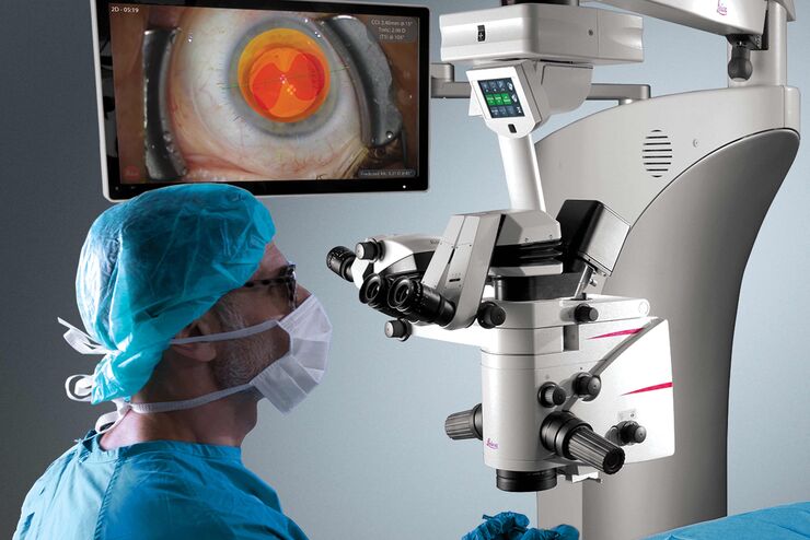
使用Proveo 8眼科手术显微镜实施的白内障与青光眼手术
这些视频录制于用Proveo 8眼科手术显微镜实施的白内障和青光眼手术期间,证明了Ike Ahmed博士在超声乳化手术中如何受益于该产品独有的CoAx 4 同轴照明技术和FusionOptics技术,CoAx 4 同轴照明技术提供了稳定的红光反射,FusionOptics技术具有能带来细节丰富的成像的高分辨率和大景深功能。