
Loading...

A Meta-cancer Analysis of the Tumor Spatial Microenvironment
Learn how clustering analysis of Cell DIVE datasets in Aivia can be used to understand tissue-specific and pan-cancer mechanisms of cancer progression
Loading...
 tissue.")
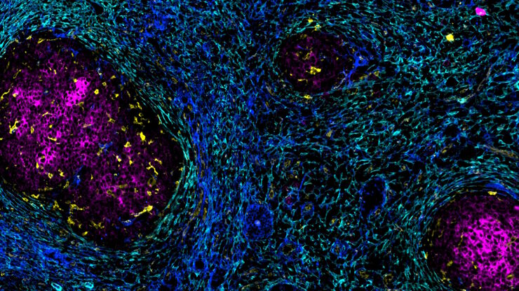
Mapping the Landscape of Colorectal Adenocarcinoma with Imaging and AI
Discover deep insights in colon adenocarcinoma and other immuno-oncology realms through the potent combination of multiplexed imaging of Cell DIVE and Aivia AI-based image analysis
Loading...

Spatial Architecture of Tumor and Immune Cells in Tumor Tissues
Dig deep into the spatial biology of cancer progression and mouse immune-oncology in this poster, and learn how tumor metabolism can effect immune cell function.
Loading...

IBEX, Cell DIVE, and RNA-Seq: A Multi-omics Approach to Follicular Lymphoma
In a recent study by Radtke et al., a multi-omics spatial biology approach helps shed light on early relapsing lymphoma patients
Loading...
Accelerating Discovery for Multiplexed Imaging of Diverse Tissues
Explore IBEX: Open-source multiplexed imaging. Join the collaborative IBEX Imaging Community for optimized tissue processing, antibody selection, and human atlas construction.
Loading...

Transforming Multiplexed 2D Data into Spatial Insights Guided by AI
Aivia 13 handles large 2D images and enables researchers to obtain deep insights into microenvironment surrounding their phenotypes with millions of detected objects and automatic clustering up to 30…
![[Translate to chinese:] Hepatocellular Carcinoma with 13 biomarkers shown – Beta-Catenin, CD3D, CD4, CD8a, CD31, CD44, CD163, DAPI, PanCK, PCK26, PD1, SMA, and Vimentin.](/fileadmin/_processed_/5/b/csm_Hepatocellular_Carcinoma_13_Markers_Zoom2_bc27c21cf2.jpg "[Translate to chinese:] Hepatocellular Carcinoma with 13 biomarkers shown – Beta-Catenin, CD3D, CD4, CD8a, CD31, CD44, CD163, DAPI, PanCK, PCK26, PD1, SMA, and Vimentin.")
Loading...
![[Translate to chinese:] Adult human Alzheimer’s brain demonstrating a panel of 15 markers.](/fileadmin/_processed_/4/9/csm_Adult_human_Alzheimers_brain_68bacde06e.jpg "[Translate to chinese:] Adult human Alzheimer’s brain demonstrating a panel of 15 markers.")
大脑的形状:阿尔茨海默病的空间生物学
阿尔茨海默病(AD)是一种神经退行性疾病,也是导致晚年认知障碍的常见原因。阿尔茨海默病的特征是出现含β-淀粉样蛋白的斑块和含磷酸化 tau 的神经纤维缠结。目前尚缺乏治疗和预防AD的有效疗法。我们将Cell DIVE与Cell Signaling Technology的抗体结合使用,检查了AD的突触过程并从空间上确定了神经胶质细胞和神经元等细胞,证明了超多标免疫荧光成像技术可用于探测AD大脑。
Loading...
![[Translate to chinese:] Co-detection of 10 extracellular matrix proteins and 3 topographical tissue landmarks by multiplex immunostaining within a single high-grade fibrous hotspot from a human hepatocellular carcinoma](/fileadmin/_processed_/0/8/csm_Single_high-grade_fibrous_hotspot_from_a_human_hepatocellular_carcinoma_e5541282bd.jpg "[Translate to chinese:] Co-detection of 10 extracellular matrix proteins and 3 topographical tissue landmarks by multiplex immunostaining within a single high-grade fibrous hotspot from a human hepatocellular carcinoma")
肝细胞癌中癌症干细胞位点的原位鉴定
在这里,我们探索了一种突破性的多重免疫检测方法,通过多重成像对细胞外基质(ECM)特征进行原位定位,从而识别肝细胞癌(HCC)内的癌症干细胞龛。