
Loading...
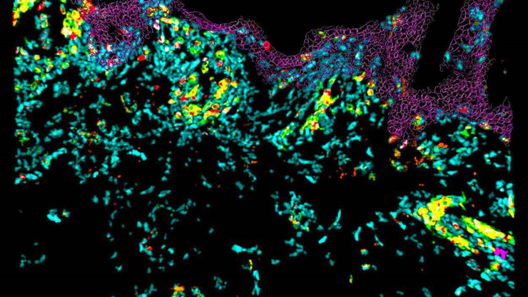
![[Translate to chinese:] Pancreatic Ductal Adenocarcinoma with 11 Apoptosis biomarkers shown – BAK, BAX, BCL2, BCLXL, Caspase9, CIAP1, NaKATPase, PCK26, SMAC, Vimentin, and XIAP.](/fileadmin/academy/2023/Gated_content/Pancreatic_Ductal_Adenocarcinoma_11_Apoptosis_Markers_ROI5.jpg "[Translate to chinese:] Pancreatic Ductal Adenocarcinoma with 11 Apoptosis biomarkers shown – BAK, BAX, BCL2, BCLXL, Caspase9, CIAP1, NaKATPase, PCK26, SMAC, Vimentin, and XIAP.")
与卢克-加蒙(Luke Gammon)一起多重成像:推进您的空间生物学研究
多重成像是一种功能强大的技术,可让研究人员同时观察单个样本中的多个目标。这对于研究复杂的生物系统尤为重要,可以帮助研究人员更好地了解不同分子和途径之间是如何相互作用的。
Loading...

Spatial Biology: Learning the Landscape
Spatial Biology: Understanding the organization and interaction of molecules, cells, and tissues in their native spatial context
![[Translate to chinese:] Cell counts for each biomarker were divided by total number of cells to give a percentage of biomarker positive cells out of total cells for each biomarker.](/fileadmin/_processed_/5/3/csm_Cell_counts_for_biomarkers_with_Cell_DIVE_ee648b397a.jpg "[Translate to chinese:] Cell counts for each biomarker were divided by total number of cells to give a percentage of biomarker positive cells out of total cells for each biomarker.")
Loading...
![[Translate to chinese:] PDAC Multiplexed imaging of CST panels enables an examination of immune cell components in pancreatic ductal adenocarcinoma (IPDAC) tissue on a single slide.](/fileadmin/_processed_/a/d/csm_Pancreatic_ductal_adenocarcinoma_tissue_3d61c18a2d.jpg "[Translate to chinese:] PDAC Multiplexed imaging of CST panels enables an examination of immune cell components in pancreatic ductal adenocarcinoma (IPDAC) tissue on a single slide.")
表征肿瘤环境以揭示洞察和空间分辨率
肿瘤环境的表征可以为癌症进展和潜在治疗靶点提供更深入的见解。我们已经使用来自Cell Signaling Technology(CST)的各种IHC验证抗体,在胰腺癌的Cell DIVE研究中验证了30多种偶联抗体。
![[Translate to chinese:] How is microscopy used in spatial biology - Teaserimage](/fileadmin/_processed_/0/f/csm_spatial-biology-ebook-tease_5ac6218fa3.jpg "[Translate to chinese:] How is microscopy used in spatial biology? A microscopy guide.")
Loading...

Multiplexing through Spectral Separation of 11 Colors
Fluorescence microscopy is a fundamental tool for life science research that has evolved and matured together with the development of multicolor labeling strategies in cells tissues and model…
Loading...
![[Translate to chinese:] Pancreatic ductal adenocarcinoma tissue section imaged with Cell DIVE](/fileadmin/_processed_/f/8/csm_Pancreatic_ductal_adenocarcinoma_tissue_section_teaser_eae5765765.jpg "[Translate to chinese:] Pancreatic ductal adenocarcinoma tissue section imaged with Cell DIVE")
多重成像的类型、优势和应用
与传统显微镜相比,多重成像技术能观察到更多的生物标记物,是一种新兴的、令人兴奋的从人体组织样本中提取信息的方法。通过同时观察多种生物标记物,可以协同探索以前只能单独探索的生物通路,并识别和探测复杂的组织和细胞表型。目前已有许多不同的多重成像方法,每种方法都采用不同的方法来实现更高的复杂性。
Loading...
![[Translate to chinese:] Colon adenocarcinoma with 13 biomarkers shown](/fileadmin/_processed_/4/9/csm_Colon_adenocarcinoma_with_13_biomarkers_shown_42edcfed0a.jpg "[Translate to chinese:] Colon adenocarcinoma with 13 biomarkers shown")
利用Cell DIVE 在单细胞水平上进行超复杂癌症组织分析
能够研究淋巴瘤细胞的异质性如何受到细胞对其微环境反应的影响,尤其是在突变、转录组和蛋白质水平上。蛋白质表达研究提供了有关细胞相互作用性质和蛋白质表达水平的最相关信息。超复合工作流程可用于研究同一癌症组织中的多种蛋白质。