
But what is spatial biology, and how can researches leverage its tools and mindset to answer the ever-increasing demands of biological questions in the post-omics era? In this article, we will provide a brief overview of the topic of spatial biology, its technologies, and products, as well as the questions that a researcher might ask that might provide access to this constantly evolving landscape.
What is spatial biology?
Put simply, spatial biology is the combination of spatial data with information about the identity of biological molecules. In other words, when XY (or XYZ) coordinate information is assigned to identified biological molecules in a sample, the resultant data may be referred to as spatial biology. However, the term is usually only invoked when performed at scale—when thousands or millions of cells, proteins, transcripts, or other analyte, are resolved spatially.
For example, while a study of a single protein and its location within a cell culture might not be called a spatial biology study, exploring that single protein in the context of a heterogeneous tissue sample among a hundred other proteins and RNA molecules is more likely to be. This allows a researcher to explore the landscape of various biological analytes in the context of the native tissue architecture and cellular microenvironment. Spatial biology can thus reveal the diversity and spatial heterogeneity of cellular biology in a tissue alongside the spatial organization of cells.
How do I generate spatial biology data?
The three main technology families that yield spatial biology data are fluorescence microscopy, sequencing, mass spectrometry, and often a combination of all three. Specific spatial biology subtypes are often defined by the base technology that positional data is added to. For example, while processing organ tissue in bulk and sequencing RNA molecules from the lysates or using single-cell sequencing could be called transcriptomics, using microdissection techniques to assign RNA sequencing reads to specific physical locations is more likely to be referred to as spatial transcriptomics or spatially-resolved transcriptomics. Spatial transcriptomics approaches can also be achieved by hybridizing barcoded tags to RNA molecules in a sample and using direct next generation sequencing or fluorescent probe reading to uncover the identity of the labeled species. Alternatively, DNA in-situ sequencing based approaches can also be adapted to the spatial biology toolset in a method known as spatial genomics. This can be used to explore, for example, copy number variations in tumor cells, or changes in chromosome counts in tissues undergoing aneuploidy—a deviation from normal chromosome quantities.
Using mass spectrometry to identify the proteins in that sample would be referred to as spatial proteomics or spatially-resolved proteomics. For this method, regions of tissue can be microdissected and subjected to mass spectrometry before those regions are realigned back to a reference image. Alternatively, various techniques for imaging mass cytometry allow regions of tissue to be directly analyzed. These techniques share a basic approach—samples are prepared and then regions of the tissue are ionizied and mass spectra is obtained and analyzed, before being related back to that spectra’s position within the sample.
This same family of techniques can also yield quantitative spatial information on non-protein metabolites, commonly referred to as spatial metabolomics, although the analytes do not necessarily have to be direct products of cellular metabolism. These types of approaches can analyze lipids (lipidomics), sugars (glycomics), or even drug molecules to assess pharmacodynamics. This is a challenging approach that relies heavily on good measurement standards but opens the door to an incredibly broad and deep understanding of the chemistry of human tissue.
Multiplexed imaging for spatial proteomics
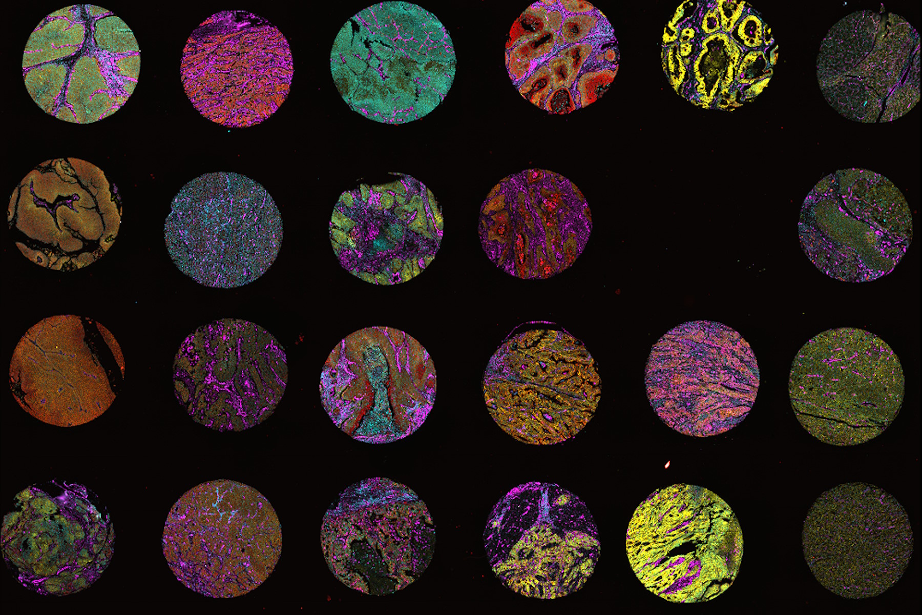
Spatial proteomics does not have to be achieved solely using mass spectrometry. It can also be accomplished using light microscopy and multiplexed imaging workflows to examine tens or hundreds of proteins in a sample. The mass spectrometry or multiplexed imaging approach are said to differ in terms of plexity, that is, the number of analytes that can be analyzed in a single sample. Looking at few molecules might be called low, or mid-plex, while looking at hundreds or thousands of molecules might be called high-plex. Why would a researcher use a lower plex technique in a spatial biology experiment? Imaging based approaches have the advantage of often providing higher resolution of imaged tissues. If knowledge of an analytes precise location within a single cell is important, multiplexed imaging might be the only avenue to achieve that result.
For example, immuno-oncology researchers are often interested in the precise cell type identities of highly specialized immune cell subtypes. This might require single cell resolution of five to ten biomarkers for an accurate phenotype to be assigned, thus requiring a higher resolution, lower-plex approach. Low/mid-plex techniques and higher plex techniques are not mutually exclusive. Lower plex approaches can define hypotheses and even specific regions of tissue to be analyzed using a mass spectrometry- or sequencing-based high-plex method later in the workflow.
How does multiplexed imaging provide an avenue for spatial biology research? There are many workflows in multiplexed spatial biology which use a variety of technologies such as:
- Staining and imaging in one
- One-pass lower-plex
- All-in-one omics solutions
- Iterative staining
For a deeper treatment of these technologies, see Multiplexed Imaging Types, Benefits and Applications.
Application example: Tumor microenvironments
Why would a researcher employ these techniques? The jump to spatial approaches comes out of a union of technological progress and a deeper knowledge of the biological activities of cells and tissues. As it becomes easier to understand what transcripts, proteins, and molecules are within cells, researchers are growing curious about how those molecules are organized, particularly within heterogenous tissues and during disease states. Consider the tumor microenvironment: a substantial amount of knowledge has revealed many of the molecular determinants of oncogenesis and cancer progression. Because of this knowledge, many classic oncogenes and oncogenetic pathways have been targeted by therapies, with mixed success. One therapeutic area with promise is that of immune therapies, which stimulate the patient’s immune system to attack and destroy tumor tissues. However, determining which patients will benefit from often highly expensive drugs can be a challenge.
The key to predicting the response to an immunotherapeutic drug is often hidden in the organization of tissues and the identity of cells in close proximity to the tumor itself. How close are tumor cells to blood vessels? Is there a presence or absence of immune cells that distinguishes the tumor from its surroundings? Are those immune cells entering or exiting the tumor area? Are they primed to respond to tumors or are the cells in an exhausted state? Does the tumor display heterogeneity in gene and protein expression, showing a differentiation into various metabolic segments? All of these questions can be relevant for predicting immunotherapy response and are perfect questions for a spatial biology approach.
Application example: Genetics and development
Classical cancer genetics may also be impacted by spatial biology techniques. A researcher might ask: where are cells in which defective DNA repair is occurring? Which cellular or genotoxic insults create these cells, and what spatial pattern of damage do particular genotoxins create? There are non-cancer, non-translational avenues for spatial biology as well. For example, complex, developing tissues such as the hair follicle display a stem cells with often quite granular cellular identities that move into different niches depending on the progress of the hair life cycle. These types of developmental biology settings are well-positioned to take advantage of spatial biology approaches. Indeed, any experimental setting that requires fine-scale knowledge of cellular identity and position can be benefited from spatial biology technologies.
Conclusion
The spatial biology landscape represents a family of tools, methods, and modes of analysis that aim to use the confluence of omics technologies and imaging to gain a deeper understanding into how human tissues are organized. As these technologies continue to advance, the scope of questions researchers can ask and answer will only continue to increase in sophistication. Complex areas of cancer biology, medicine, developmental biology, and beyond stand poised to be deeply impacted by these technological breakthroughs. Adopting the “mindset” of spatial biology will help ease the transition to using these methods, and encourage researchers to think about how this approach might provide new insights into their current experimental systems and beyond.
