
Loading...

Find Relevant Specimen Details from Overviews
Switch from searching image by image to seeing the full overview of samples quickly and identifying the important specimen details instantly with confocal microscopy. Use that knowledge to set up…
![[Translate to chinese:] AiviaMotion: Truly simultaneous multicolor imaging of live cells (U2OS) in 3D](/fileadmin/_processed_/b/1/csm_How_Artificial_Intelligence_Enhances_Confocal_Imaging_teaser_52a5f9637d.jpg "[Translate to chinese:] AiviaMotion: Truly simultaneous multicolor imaging of live cells (U2OS) in 3D")
Loading...
 in 3D")
How Artificial Intelligence Enhances Confocal Imaging
In this article, we show how artificial intelligence (AI) can enhance your imaging experiments. Namely, how Dynamic Signal Enhancement powered by Aivia improves image quality while capturing the…
Loading...
Spectroscopic Evaluation of Red Blood Cells
Hemoglobinopathies are a major healthcare problem. This study presents a possible diagnostic tool for thalassemia which is based on confocal spectroscopy. This approach exploits spectral detection and…
Loading...
Visualizing Protein Degradation and Aggregation in the Living Cell
Our guest speaker, Prof Dr Eric Reits, presents his work on neurodegenerative disorders. Reits’ group are experts on the subject of Huntington’s disease and work towards identifying leads for…
Loading...
Life Beyond the Pixels: Deep Learning Methods for Single Cell Analysis
Our guest speaker Prof Dr Peter Horvath presents his work on single cell-based large-scale microscopy experiments. This novel targeting approach includes the use of machine learning models and…
Loading...
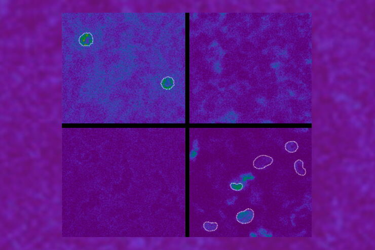
超分辨率显微镜图片库
由于光的衍射极限,传统共聚焦显微镜无法分辨约240纳米以下的结构。当需要提高分辨率以研究衍射极限尺度以下的结构和分子事件时,会使用超分辨率显微镜技术,如STED、PALM或STORM,或某些解卷积处理方法。
Loading...
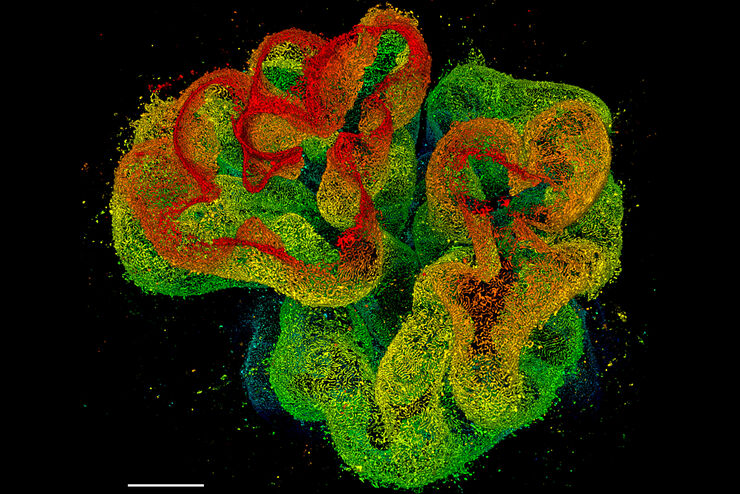
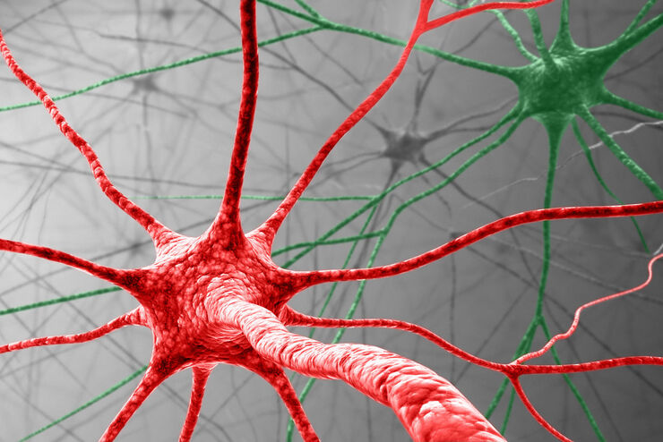
组织图片库
对动物和人体组织进行视觉分析对于了解癌症或神经变性等复杂疾病至关重要。从基本的免疫组化到体内成像,共聚焦显微镜和先进的模式可以让人们了解细胞、生物分子及其在环境中的相互作用。
Loading...
![[Translate to chinese:] Nematostella](/fileadmin/_processed_/d/c/csm_Nematostella-LiveImaging-Stellaris_1d96dd4af5.jpg "[Translate to chinese:] Nematostella")
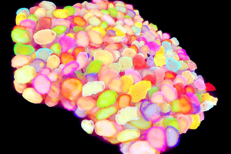
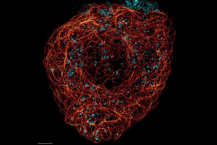
活细胞成像图库
活细胞显微镜技术是更好地了解细胞和分子功能的基础。如今,宽场显微镜是用于长时间观察细胞动态和发育的最常用技术。共聚焦显微镜也是一种重要工具,可生成三维结构图像,并以高空间和时间分辨率研究高度动态的细胞过程,同时使标本保持接近原生状态。