
Here are some reasons why users can rapidly obtain a complete overview of their specimens to find regions of interest (ROIs) and then easily acquire high-resolution images with the LAS X Navigator software.
Get a quick orientation of the sample
Finding nicely labelled cells on a Petri dish or a particular ROI in a large brain tissue section can be difficult and time consuming. The reason is that many biological samples are much larger than the maximum field of view of the microscope, making it troublesome for users to find where they are on the specimen.
It is possible to quickly create an overview of the sample for selecting the ROIs for further investigation with the spiral-scan function of LAS X Navigator. It allows users with a single click to acquire a mosaic scan which is centered around the current field of view.
Work seamlessly between different size scales
While users want to get the overall sample, they are also interested in seeing the finest specimen details. This means you must seamlessly switch between different size scales.
Users can swiftly change between different objectives with the LAS X Navigator software and scan the sample with the appropriate level of detail.
When the objective lens is changed, all relevant parameters related to tile size and position are automatically recalculated. Moreover, the viewer function can display the overlay of multiple recordings, including those acquired with different objective lenses. This way, users can seamlessly probe the sample at different scales by first acquiring a quick overview, then seeing details from multiple, arbitrary-shaped ROIs using high resolution z-stacks.
Stay in-focus on large samples and during time-lapse recordings
Users may need to image a large sample, such as a tissue section, in its entirety or scan a multiwell plate. When performing these kinds of experiments, there is often the disadvantage that biological samples and probe carriers are not perfectly flat. These geometric imperfections, as well as sample drift, during time-lapse recordings both lead to out-of-focus problems during time-lapse recordings.
There are multiple ways to compensate for these issues with the LAS X Navigator software. It has an intuitive way for creating a so-called “focus map” by defining manually or automatically an arbitrary number of focus points for interpolation of the appropriate z position. Moreover, as the “Adaptive Focus Control” (AFC) is fully integrated into the software, a non-invasive, fast approach is available for keeping the sample in focus over an extended period of time.

Assay editor and statistical sampling
When users want to probe several manipulations at once and still get statistically meaningful results, the natural choice is to work with a multiwell carrier for higher throughput. Setting up these kinds of experiments manually is tedious and limits productivity, because for every single well multiple imaging positions must be acquired by hand.
Using the Assay Editor (license separately available) of the LAS X Navigator software, a wide selection of common probe carriers is ready for use, such as slides, dishes, and multiwell plates. Users simply choose the correct carrier and select how they want to statistically sample each well. This way of doing it allows a fast setup so the focus is on obtaining meaningful results. Instead of using a standard carrier, users can customize an individual carrier.

Predefined Navigator protocols can speed up things further. In the following example, a protocol for an 8-chamber slide with 3 positions in each well was loaded and executed using a DMi8 widefield system equipped with a Quantum stage.
Integrated image stitching and analysis
Acquiring high-quality data is important, but the experiment is not quite done at that point. Users still need to stitch the single tile scans and perform the image analysis. To this end, the LAS X Navigator software facilitates such workflows: After image acquisition, there is the option to automatically merge single tiles using the selected stitching algorithm. Also, the processing functionality of the 3D Analysis module (license separately available) is fully integrated into the software to support the entire workflow. For example, a processing pipeline can be used to characterize cell nuclei which were just recorded, i.e., segment them, count them, measure their size, etc.

Image fusion to integrate information from external images
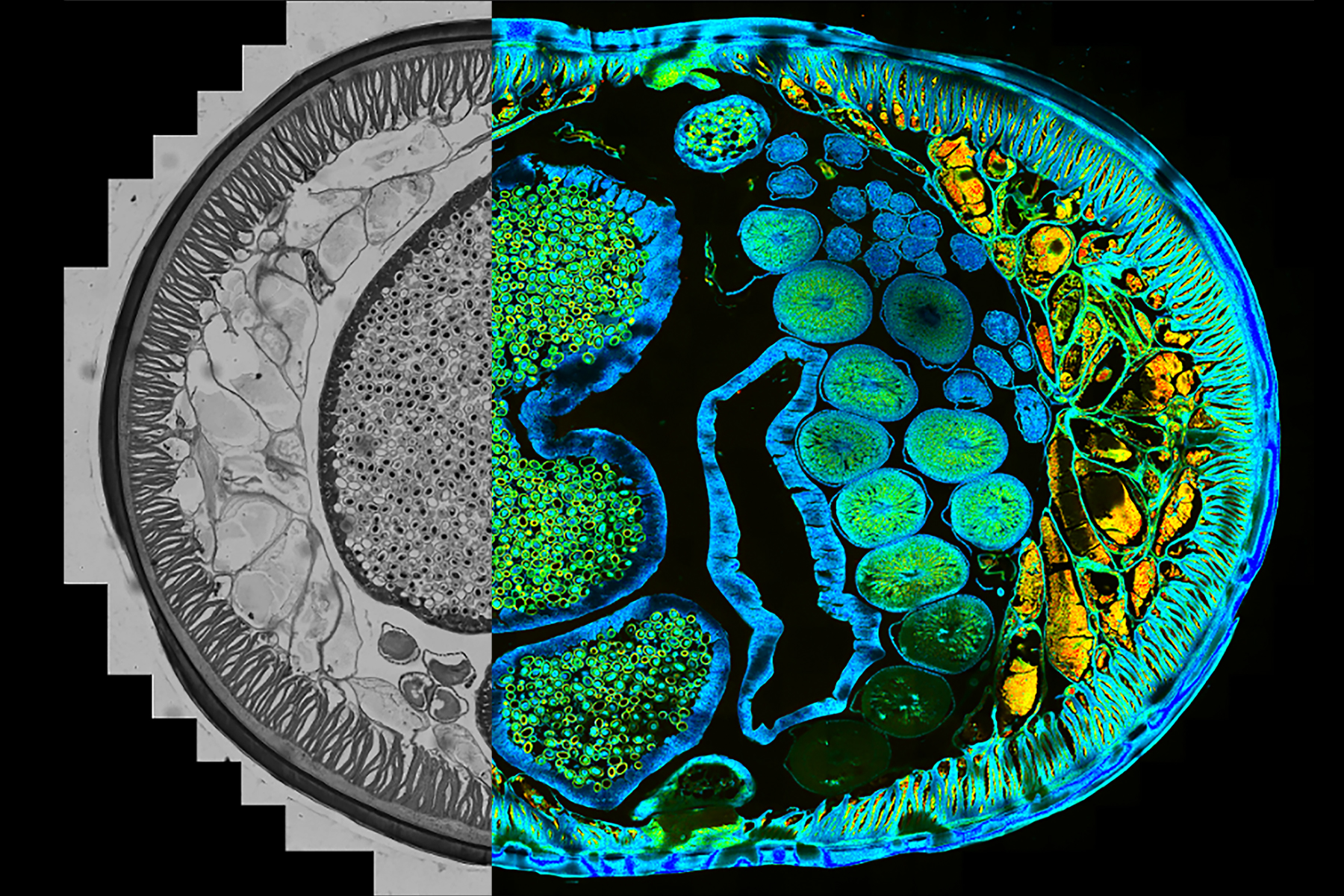
What if a pre-scan of the sample was already done with another system, e.g., a Leica widefield microscope, and now the high-resolution scan needs to be done with a STELLARIS confocal microscope? Naturally, the users want to combine these imaging data. Images from different sources can be imported and aligned with the LAS X Navigator software.
Users simply select the images they want to import into the software and use its toolbox to swiftly align the images. This way, even a snapshot from a smartphone camera could be used as a bird’s eye view for defining ROIs and scanning them automatically.

Integrate fluorescence lifetime-based imaging with STELLARIS (TauSense and FALCON)
At times, users need to separate spectrally overlapping fluorophores, reduce unwanted contributions to the signal or study micro-environmental changes. While intensity-based imaging information is still the most used dimension for fluorescence microscopy, many have learned to appreciate fluorescence lifetime-based imaging.
LAS X Navigator allows users to acquire fluorescence lifetime data with a stage application.
The TauSense tools are available with LAS X Navigator to recorddifferences in the average arrival time using TauContrast or distinguish certain spectrally overlapping fluorophores by employing TauSeparation. Also, FALCON is fully compatible with LAS X Navigator.

 and phalloidin (magenta), imaged using Viventis SCAPE; scale bar 50μm. Courtesy of Marina Cuenca and Heleen Jungen (Dayton lab), EMBL Barcelona.")


