Loading...
![[Translate to chinese:] Living HeLa cells stained with WGA-488 (yellow), SPY-Actin (cyan), and SiR-Tubulin (magenta). Instant Computational Clearing (ICC) was applied. [Translate to chinese:] Living HeLa cells stained with WGA-488 (yellow), SPY-Actin (cyan), and SiR-Tubulin (magenta). Instant Computational Clearing (ICC) was applied.](/fileadmin/_processed_/b/0/csm_Living_HeLa_cells_Instant_Computational_Clearing_e2a2820021.jpg)
如何进行动态多色延时成像
本文将举例说明 Mica 进行动态活细胞成像的能力。活细胞成像揭示了各种细胞事件。由于其中许多事件具有快速动态性,显微镜成像系统必须足够快才能记录下每一个细节。这种成像系统的一个主要优势是能够同时捕获多个荧光成像通道,以精确地显示它们的时空相关性。
Loading...
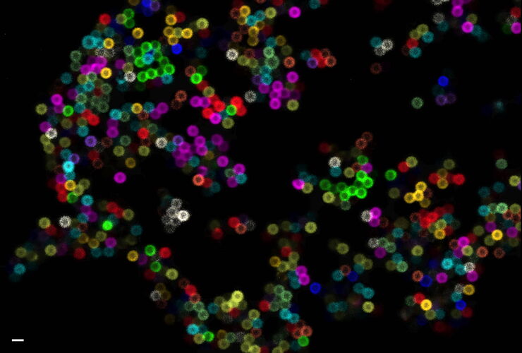
Multiplexing through Spectral Separation of 11 Colors
Fluorescence microscopy is a fundamental tool for life science research that has evolved and matured together with the development of multicolor labeling strategies in cells tissues and model…
Loading...
![[Translate to chinese:] Projection of a confocal z-stack. Sum159 cells, human breast cancer cells [Translate to chinese:] Projection of a confocal z-stack. Sum159 cells, human breast cancer cells kindly provided by Ievgeniia Zagoriy, Mahamid Group, EMBL Heidelberg, Germany. Blue–Hoechst - indicates nuclei, Green–MitoTracker mitochondria, and red–Bodipy - lipid droplets](/fileadmin/_processed_/6/6/csm_Keyvisual-Cancer-cell-under-Cryo_Coral-Cryo_TechNote_99f1c3cd0c.jpg)
低温光学显微镜的新成像工具
荧光显微镜图像能够为cryo-FIB加工提供定位支持,其质量决定了所制备薄片的结果。本文描述了LIGHTNING技术是如何显著提高图像质量,以及如何利用该技术基于荧光寿命的信息来辨别样品的不同结构。
Loading...
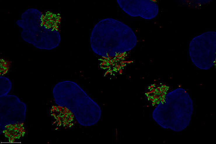
高尔基组织对细胞应激的反应变化
在本集MicaCam直播活动中,来自海德堡欧洲分子生物学实验室的特邀嘉宾George Galea将对用各类化疗药物进行治疗的HeLa Kyoto细胞进行分析,并观察其对高尔基复合体和细胞核的组织和定位的影响。
Loading...
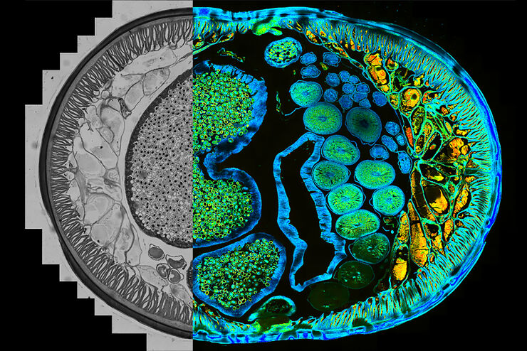
Find Relevant Specimen Details from Overviews
Switch from searching image by image to seeing the full overview of samples quickly and identifying the important specimen details instantly with confocal microscopy. Use that knowledge to set up…
Loading...
![[Translate to chinese:] LNG-non-LNGHeLa cells. Cells kindly provided by I. Zagoriy, Mahamid Group, EMBL Heidelberg [Translate to chinese:] LNG-non-LNGHeLa cells labeled with light blue –Hoechst, Nuclei](/fileadmin/_processed_/c/6/csm_LNG-non-LNGHeLa-cells_e052df4846.jpg)
精确三维定位,实现EM成像——掌握精髓
低温电子断层扫描(CryoET)是一种成像技术,可以让研究人员以亚纳米分辨率观察蛋白质和其他大生物分子。了解分子的形状和结构,包括口袋和裂隙,可以帮助研究人员设计能够像拼图一样附着于分子的药物。低温ET成像也因此成为了解和治疗疾病和失调的重要基础。
Loading...
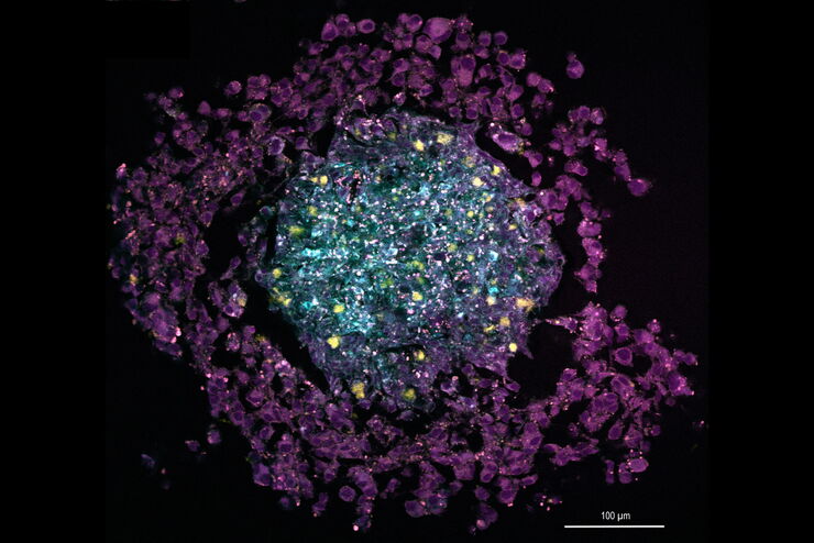
The Potential of Coherent Raman Scattering Microscopy at a Glance
Coherent Raman scattering microscopy (CRS) is a powerful approach for label-free, chemically specific imaging. It is based on the characteristic intrinsic vibrational contrast of molecules in the…
Loading...
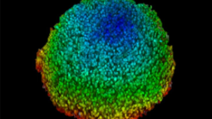
Imaging of Anti-Cancer Drug Uptake in Spheroids using DLS
Spheroid 3D cell culture models mimic the physiology and functions of living tissues making them a useful tool to study tumor morphology and screen anti-cancer drugs. The drug AZD2014 is a recognized…

![[Translate to chinese:] Zebrafish heart showing the ventricle with an injury in the lower area [Translate to chinese:] Zebrafish heart showing the ventricle with an injury in the lower area](/fileadmin/_processed_/9/6/csm_Zebrafish_heart_showing_ventricle_with_injury_teaser_490e470f4e.jpg)