
![[Translate to chinese:] Zebrafish heart showing the ventricle with an injury in the lower area](/fileadmin/_processed_/9/6/csm_Zebrafish_heart_showing_ventricle_with_injury_teaser_490e470f4e.jpg "[Translate to chinese:] Zebrafish heart showing the ventricle with an injury in the lower area")
Loading...
")
Wt1 Genes Can Induce a Cardiomyocyte to Epicardial-like Cell Fate Transition
From this study, it was concluded that Wt1 plays a yet undescribed role for cardiomyocyte differentiation by repressing chromatin opening at specific genomic loci and that sustained ectopic expression…
Loading...
, unsaturated lipids (magenta, 3050 cm-1), collagen (SHG, cyan). Sample courtesy of R. Rudolf, J Klicks, Hochschule Mannheim")
The Potential of Coherent Raman Scattering Microscopy at a Glance
Coherent Raman scattering microscopy (CRS) is a powerful approach for label-free, chemically specific imaging. It is based on the characteristic intrinsic vibrational contrast of molecules in the…
Loading...
A Versatile Palette of Fluorescent Probes
Researchers at the Max Planck Institute for Medical Research in Heidelberg have developed a general strategy to synthesize live-cell compatible fluorogenic probes, and the result are the new MaP (Max…
Loading...
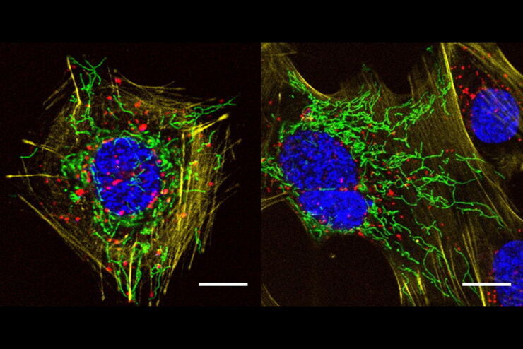
如何为免疫荧光显微镜制备样本
免疫荧光(IF)是一种用于可视化观察细胞内过程、状态和结构的强大工具。IF制剂可通过多种显微镜技术(如激光共聚焦、宽场荧光、全内反射成像等)来加以分析,具体取决于应用目的或研究人员的关注重点。与此同时,在很多使用至少一套简易荧光显微镜的研究工作组当中,IF早已成为不可缺少的一部分。
Loading...

Imaging of Anti-Cancer Drug Uptake in Spheroids using DLS
Spheroid 3D cell culture models mimic the physiology and functions of living tissues making them a useful tool to study tumor morphology and screen anti-cancer drugs. The drug AZD2014 is a recognized…
Loading...

Intravital Microscopy of Cancer
Join our guest speaker Prof Dr Jacco van Rheenen, as he presents his work on the identity, behavior and fate of cells that drive the initiation and progression of cancer.
![[Translate to chinese:] AiviaMotion: Truly simultaneous multicolor imaging of live cells (U2OS) in 3D](/fileadmin/_processed_/b/1/csm_How_Artificial_Intelligence_Enhances_Confocal_Imaging_teaser_52a5f9637d.jpg "[Translate to chinese:] AiviaMotion: Truly simultaneous multicolor imaging of live cells (U2OS) in 3D")