
Loading...
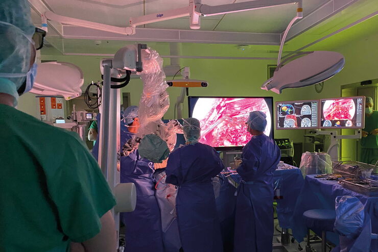
Optimal Visualization in Brain Surgery
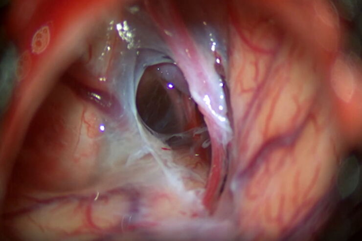
This case study “Treatment of the Galassi type III arachnoid cyst with the M530 OHX surgical microscope from Leica Microsystems” documents the procedure step by step and shows the visualization…
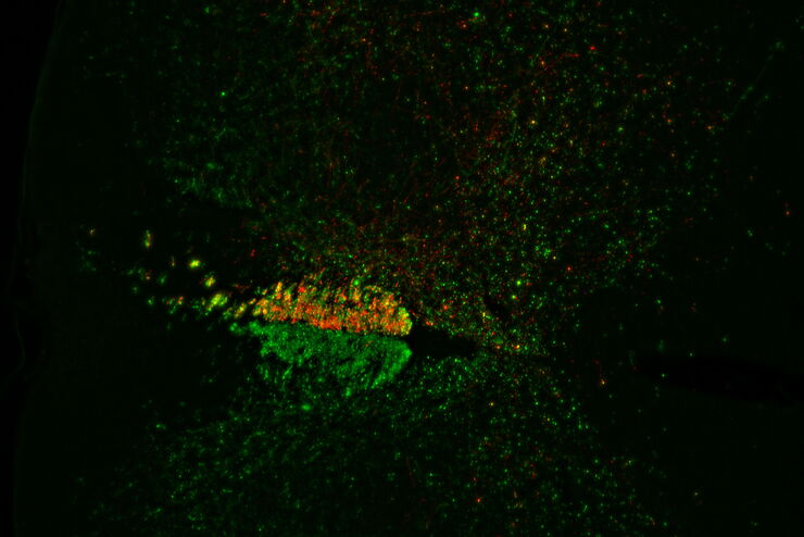
![[Translate to chinese:] Virally labeled neurons (red) and astrocytes (green) in a cortical spheroid derived from human induced pluripotent stem cells. THUNDER Model Organism Imagerwith a 2x 0.15 NA objective at 3.4x zoomwas used to produce this 425 μm Z-stack (26 positions), which is presented here as an Extended Depth of Field(EDoF)projection.](/fileadmin/_processed_/e/c/csm_THUNDER_Imager_Model-Org_Header-Gallery-Neuroscience_4652e132e6.jpg "[Translate to chinese:] Virally labeled neurons (red) and astrocytes (green) in a cortical spheroid derived from human induced pluripotent stem cells.")
Loading...
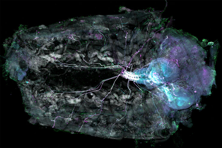
Understanding Motor Sequence Generation Across Spatiotemporal Scales
We have developed a microscopy-based pipeline to characterize a developmentally critical behavior at the pupal stage of development, called the ecdysis sequence. We study brain-wide neuronal activity…
Loading...
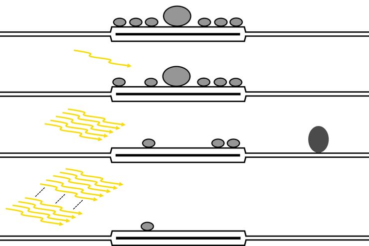
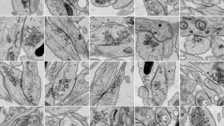
使用增强功能电子显微镜研究大脑切片中的突触
神经科学的一个基本问题就是突触的结构与其功能特性之间有何关系?过去几十年,电生理学揭示了突触传递机制,而电子显微镜(EM)深入探索了突触形态。用于关联突触生理学和超微结构的方法可以追溯到20世纪中叶。目标是获得突触传递的快照,即捕获电子显微照片中的动态过程。
Loading...
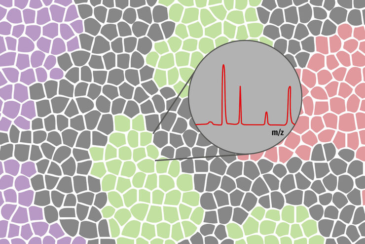
如何利用激光显微切割来改善生物标记物识别与分离
生物标记物可用作特定疾病如癌症的指征标记。这样一来,肿瘤微环境就容易引起人们的警觉。但在肿瘤区域和非肿瘤区域以及肿瘤本身之间存在着明显的分子差异。这些情况只能通过分离这些区域的特定的、微小的部分来破译。
Loading...
Neurosurgery with Heads-up Display
In the following video interviews Prof. Dr. Raphael Guzman, Vice Chairman of the Department of Neurosurgery at the University Hospital in Basel, Switzerland, talks about his experience in heads-up…
Loading...
Evaluating Axon Regeneration After Brain or Spine Trauma of Mice
Damaged nerve regeneration was investigated using mouse spinal cord sections treated with compounds that counter axon growth inhibitor (AGI) proteins. The sections were screened to find active and…
Loading...
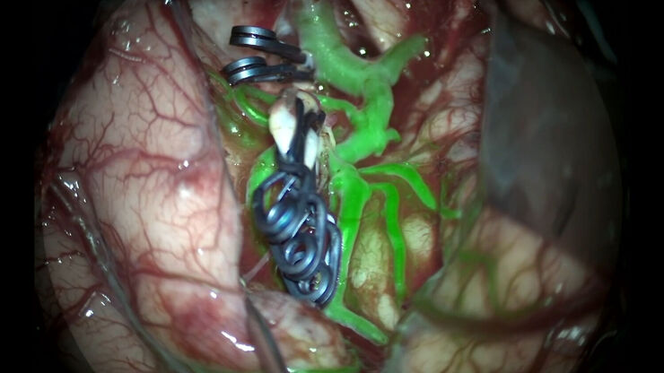
GLOW800 Augmented Reality Fluorescence in Aneurysm Treatment
This case study from Prof. Dr. Feres Chaddad talks about the treatment of unruptured MCA (middle cerebral artery) and PCOM (posterior communicating artery) aneurysms with microsurgical clipping. It…
Loading...
通过光遗传和电刺激技术研究纳米桥接结构和动力学
纳米级超微结构信息通常是由经固定和处理样品的静态图像获得的。但是,这些静态图像只是不断变化的动态结构中的一个瞬间。因此,如何探索动态过程中的特定时间点,是纳米级超微结构研究的一个重大挑战。通过光遗传或电刺激技术,并结合毫秒级样品玻璃化技术探索纳米级超微结构,是一种解决上述问题具有前景的技术。在本应用白皮书的第一部分中,我们将从实际应用角度讨论光刺激辅助的样品玻璃化工作流程。