
Loading...
![[Translate to chinese:] Mouse lung sections](/fileadmin/_processed_/c/3/csm_Studying_Pulmonary_Fibrosis_Teaser_fad792a540.jpg)
肺纤维化研究
本文中所示结果表明,相比明场,使用偏振光可以更清晰地分辨小鼠肺组织中的胶原蛋白纤维化和非纤维化区域。为更好地理解促使瘢痕组织形成的肺纤维化,正常情况下会研究组织中的纤维化区域。分别使用明场和偏振光对小鼠肺组织中的胶原蛋白纤维化病变区域进行成像。成像胶原蛋白时,使用一般的染色法和明场显微镜很难区分纤维化和非纤维化区域。本文中使用偏振光对肺组织进行成像,两个区域呈现出非常明显的颜色差异。
Loading...
控制药品中的微粒污染
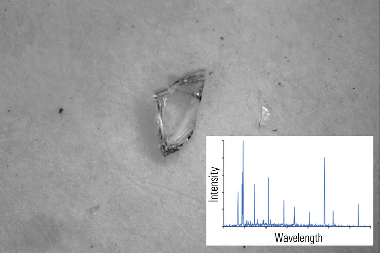
本文阐述了如何使用光学显微镜和激光诱导击穿光谱(LIBS)相结合的二合一方法识别制药行业中的微粒污染物。药物和静脉注射溶液等药品的微粒污染可能会导致严重问题。为消除药品微粒污染,最重要的是能够快速、准确地识别污染,甚至能够快速找到污染源。激光诱导击穿光谱可以对材料进行快速的多元素分析。本文介绍的二合一方法可以同时提供目视检查(颜色和形状)和化学(成分)分析,可快速、可靠地识别非监管环境中的微粒污染…
Loading...
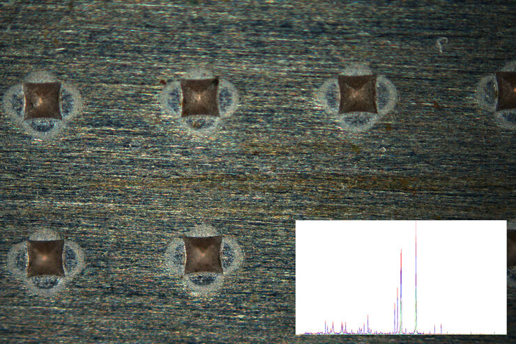
案例介绍:(USB连接器)污染物和底层基材的快速视觉和化学分析
本报告中介绍了一种视觉和化学分析二合一解决方案,可以更高效、更完整地分析材料污染物。除了同步的视觉和化学检测,还可以使用光学显微镜和激光诱导击穿光谱(LIBS)二合一解决方案,快速清除污染物并检测底层基材。
Loading...

Studying Human Brain Development and Disease
Neural spheroids created from human induced pluripotent stem cells (iPSCs) provide effective and novel tools for studying brain development, as well as the underlying pathological mechanisms of…
Loading...
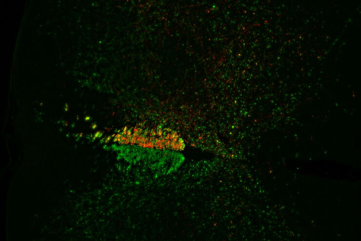
Evaluating Axon Regeneration After Brain or Spine Trauma of Mice
Damaged nerve regeneration was investigated using mouse spinal cord sections treated with compounds that counter axon growth inhibitor (AGI) proteins. The sections were screened to find active and…
Loading...
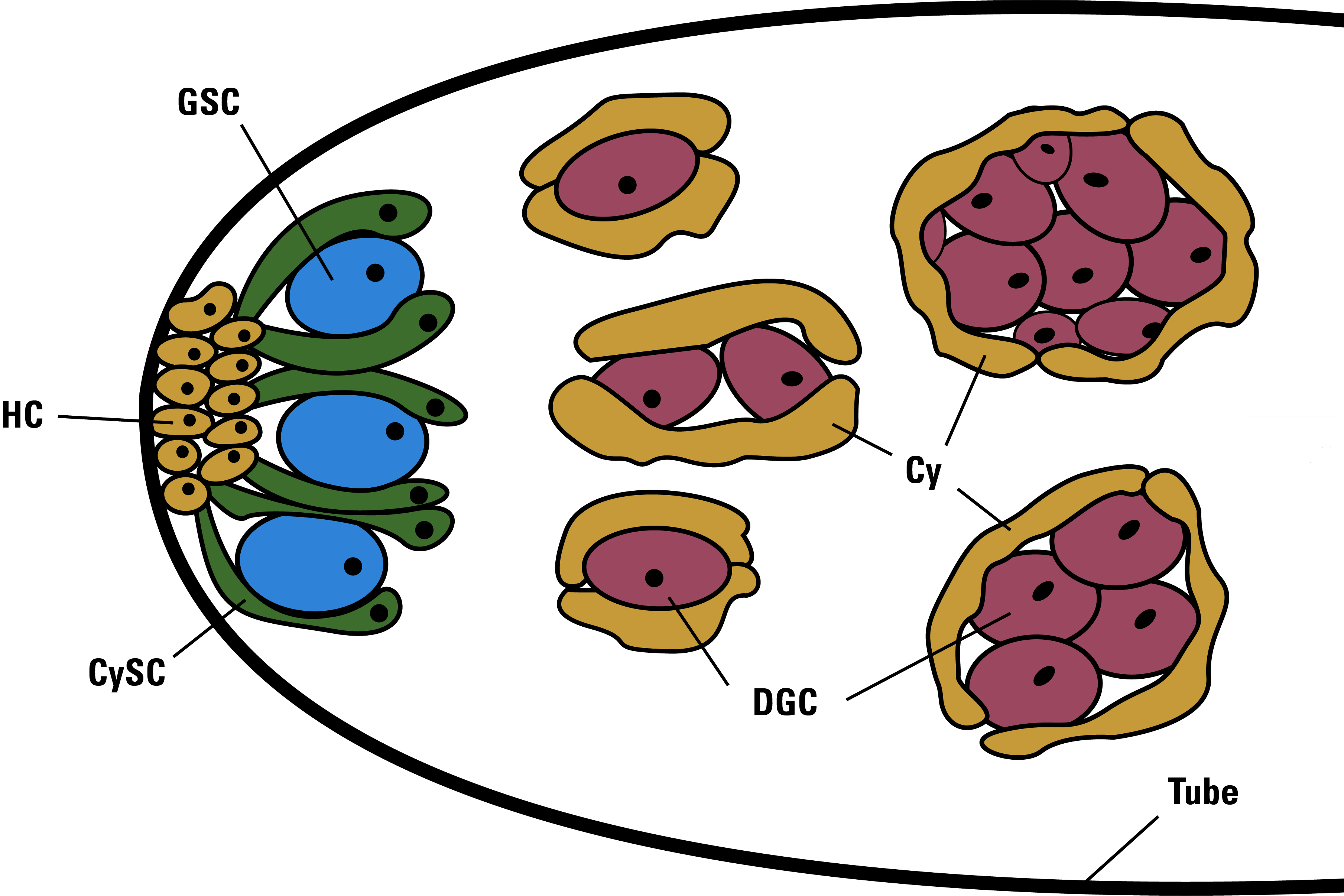
Drosophila Testis Niche Stem Cells – Three Color Computational Clearing
Differentiated living beings such as humans, but also a fruit fly or a plant, possess not only the differentiated cells which form specific tissues, but also those cells whose fate is not yet (or only…
Loading...
Alzheimer Plaques: fast Visualization in Thick Sections
More than 60% of all diagnosed cases of dementia are attributed to Alzheimer’s disease. Typical of this disease are histological alterations in the brain tissue. So far, there is no cure for this…