연락처

상관관계적 광학 현미경과 전자현미경(CLEM)

마이크로미터와 나노미터 크기 사이의 간격을 메울 수 있고, 기능적 정보와 구조적 정보를 연결할 수 있다고 상상해 보십시오. 특정 연구 질문에 대해, 이것은 살아있는 세포 이미징, 고압 동결 고정, 초박절편, (극저온) 전자현미경과 같은 여러 방식을 결합시키기 위해 다양한 시료 준비와 현미경 기법을 사용하는 것 의미합니다. CLEM 접근 방식은 이러한 격차를 해소하도록 도와드립니다.

라이카마이크로시스템즈 CLEM 솔루션은 시료 생존성, 품질 확인, 정밀하고 신뢰할 수 있는 3D 타게팅 메커니즘을 보장합니다. 사용자는 적절한 시점에 적합한 시료를 직접 확인하고 높은 분해능의 극저온 공처점 데이터를 얻거나 초미세구조 환경에 형광 정보를 넣기 위해 이 솔루션을 이용할 수 있습니다.

도움이 필요하십니까?

상관관계적 광학 현미경과 전자현미경 솔루션에 대한 라이카 전문가들이 조언으로 기꺼이 도와드립니다.

CLEM은 어디에 사용됩니까?

CLEM 접근법은 나노미터 이하 분해능으로 시료를 더 자세히 조사하는 데 사용됩니다. 한 영역에서 여러 층의 정보를 조합할 수 있을 뿐만 아니라, 어두울 때 등대가 있는 것과 같이 넓은 곳에서 관심 영역의 특정 대상을 지정할 수도 있습니다.

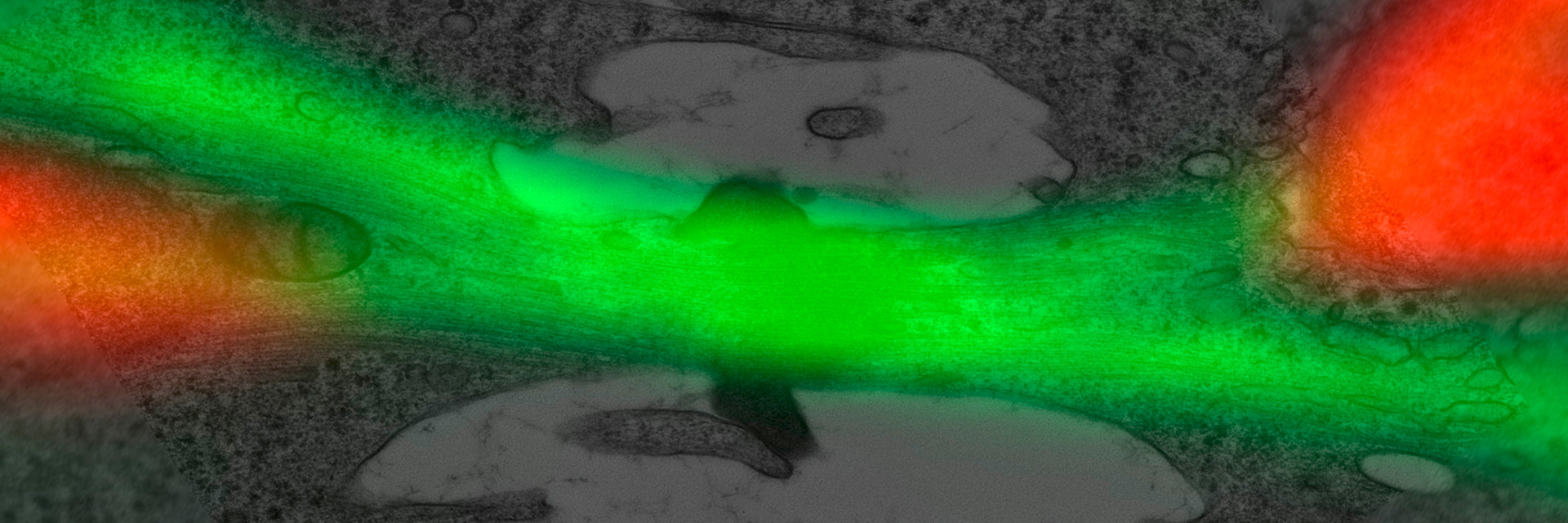
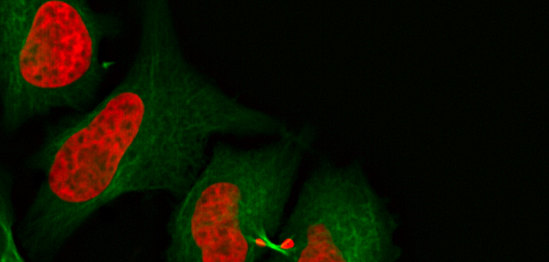
예를 들어, Cryo-EM을 사용한 단백질 구조 분석을 위해 극저온 형광 현미경으로 쉽게 표지된 단백질을 검출하고 타겟팅할 수 있습니다(Coral Cryo에 대해 자세히 알아보기). 또 다른 예는 기능적 정보를(예: 세포 간 상호작용) 구조 정보와 결합하고 광 분해능 한계 이상이 필요할 때입니다(Coral Life에 대해 자세히 알아보기).

사실 말로는 쉬워 보이지만, 성공적으로 (cryo) CLEM 작업 흐름을 만드는 데는 많은 어려움이 있습니다. 이미 실험 설계 단계에 있을 때, 사용자는 다음 질문을 다루기 위해 고려해 봐야 합니다:

  • 시료를 최적의 상태로 준비하고 유지 방법은?
  • 관심 대상 구조를 식별하고 표시하는 방법은?
  • 기기 간에 시료를 안전하고 효율적으로 운반하는 방법은?
  • 이어지는 전자현미경 단계들을 위해 적합한 형식으로 이미징하고 목표 데이터를 제공하는 방법은?

Coral 제품군은 시료를 단계적으로 처리하도록 돕고 CLEM 작업 흐름의 신뢰성을 높입니다.

CLEM 데이터를 어떻게 상호 연관시킬 수 있습니까?

광학 현미경과 전자현미경의 상관관계는 실온 실험과 cryo-CLEM 실험 모두에서 성공적인 실험을 위해 중요합니다. 좌표 조정 시스템이 설치되어 있어야 되고 다양한 이미지 방식에서 잘 인식되어야 합니다. 다음으로, 높은 정밀도로 고해상도 정보를 되찾아오기 위해 영상 맞춤(넓은 개요 데이터를 위함) 또는 위치의 절대 좌표로(x,y,z, 값) 영상 정보를 덮어 씌웁니다.

극저온 CLEM이란 무엇입니까?

Cryo-CLEM(상관관계적 광학 현미경과 전자현미경)에서는 두 이미징 단계가 극저온 조건에서 수행이 되며, 이는 신뢰할 수 있는 극저온 시료 준비 작업 흐름에 대한 필요성을 의미입니다. 여기에는 신뢰할 수 있는 시료 유리화(vitrification), 안전한 시료 운반 및 안정적인 극저온 광학 현미경 이미징이 포함됩니다. 얼음 오염에 특히 주의를 기울여야 합니다: 높은 습도 환경과 더러운 LN2는 시료를 쉽게 오염 시킬 수 있습니다. Coral Cryo 제공 사항: 1) 안전하고 깨끗한 시료 처리, 2) 안정적이고 오랜 기간 동안 극저온 현미경 이미징, 3) Coral Cryo 소프트웨어를 사용하여 본래 환경 내에서 생체 분자의 이미징 및 정밀 타게팅.

생명 과학 연구

라이카사의 생명과학사업부는 미세구조의 시각화 및 분석을 위한 혁신 기술 및 기술 전문성을 원하는 scientific community 를 충족시키는 imaging을 지원할 수있습니다. 라이키의 고객을 과학분야의 선두자로 이끌어내는것에 관심이 있습니다.

Read More

바이러스 연구

연구의 관심 분야가 바이러스 감염과 질병에 집중되어 있습니까? 라이카마이크로시스템즈의 이미징 및 샘플 준비 솔루션을 통해 바이러스학에 관한 통찰력을 얻는 방법을 알아보세요.

Read More

신경과학 연구

신경변경 질환에 대해 더 잘 이해하기 위해 노력하고 있거나 신경계 기능을 연구하고 계십니까? 라이카마이크로시스템즈의 이미지 솔루션을 통해 발전을 이룰 수 있는 방법을 알아보세요.

Read More

암 연구

암은 성장 통제에 결함이 있는 세포에 의해 발생하는 복잡하고 이질적인 질병입니다. 하나 또는 한 그룹의 세포에서 일어나는 유전적 또는 후생적 변화가 정상적인 기능을 방해하고, 자율적이고 통제되지 않는 세포 성장과 증식을 초래합니다.

Read More

관련 문서

상관관계적 광학 현미경과 전자현미경 (CLEM) 관련 최신 기사 보기

라이카마이크로시스템즈 지식 포털은 현미경에 관한 과학 연구 및 교육 자료를 제공합니다. 초보자와 숙련된 기술자, 과학자가 일상 업무와 실험에 모두 사용할 수 있는 콘텐츠로 구성되어 있습니다.

더 많은 기사
Water Flea Daphnia imaged by Electron Microscopy. Courtesy of Mag. Dr. Gruber, University of Vienna, Austria

Streamline your EM Sample Preparation Workflow for Biological Applications

Master EM sample preparation, including ultramicrotomy, for life sciences in this expert eBook!
Micrograph of dinoflagellate cells. Scale bar = 1 µm.

How Marine Microorganism Analysis can be Improved with High-pressure Freezing

In this application example we showcase the use of EM-Sample preparation with high pressure freezing, freeze substiturion and ultramicrotomy for marine biology focusing on ultrastructural analysis of…
Projection of a confocal z-stack. Sum159 cells, human breast cancer cells kindly provided by Ievgeniia Zagoriy, Mahamid Group, EMBL Heidelberg, Germany. Blue–Hoechst - indicates nuclei, Green–MitoTracker mitochondria, and red–Bodipy - lipid droplets

New Imaging Tools for Cryo-Light Microscopy

New cryo-light microscopy techniques like LIGHTNING and TauSense fluorescence lifetime-based tools reveal structures for cryo-electron microscopy.
Correlation of markers in the LM and the FIB image.

How to Target Fluorescent Structures in 3D for Cryo-FIB Milling

This article describes the major steps of the cryo-electron tomography workflow including super-resolution cryo-confocal microscopy. We describe how subcellular structures can be precisely located in…
HeLa cells labeled with dark blue – Hoechst, Nuclei; magenta – MitoTracker Green, Mitochondria; turquoise - Bodipy, lipid droplets. Cells kindly provided by Ievgeniia Zagoriy, Mahamid Group, EMBL Heidelberg, Germany.

Precise 3D Targeting for EM Imaging - Access What Matters

Find out how the seamless cryo-electron tomography workflow Coral Cryo uses confocal super resolution to target your structure of interest more precisely.

How to Successfully Perform Live-cell CLEM

The Leica Nano workflow provides a streamlined live-cell CLEM solution for getting insight bout structural changes of cellular components over time. Besides the technical handling described in the…

How to Successfully Implement Coral Life

The live-cell  CLEM workflow allows you to capture dynamic information related to a relevant biological process as it happens and put these observations into their ultrastructural context. The Leica…

Advancing Cellular Ultrastructure Research

Freeze-fracture and freeze-etching are useful tools for studying flexible membrane-associated structures such as tight junctions or the enteric glycocalyx. Freeze-fracture and etching are two…
HeLa Kyoto cells (HKF1, H2B-mCherry, alpha Tubulin, mEGFP). Left image: Maximum projection of a z-stack prior to ICC and LVCC. Right image: Maximum projection of a mosaic z-stack after ICC and LVCC.

How to Improve Live Cell Imaging with Coral Life

For live-cell CLEM applications, light microscopy imaging is a critical step for identifying the right cell in the right state at the right time. In this article, Leica experts share their insights on…

The Cryo-CLEM Journey

This article describes the Cryo-CLEM technology and the benefits it can provide for scientists. Additionally, some scientific publications are highlighted. Recent developments in cryo electron…
The EM ICE Nano loading area

How to Keep Your Samples Under Physiological Conditions

The Coral Life workflow combines dynamic data with the best possible sample fixation by high pressure freezing. However, good sample preservation won’t help if your cells are stressed by temperature…

Putting Dynamic Live Cell Data into the Ultrastructural Context

With workflow Coral Life, searching for a needle in the haystack is a thing of the past. Take advantage of correlative light and electron microscopy to identify directly the right cell at the right…
3D reconstruction of an intercellular bridge in a C. elegans embryo

Download EM Workflow Solutions Booklet

This publication is a compilation of appropriate workflows for the most frequently used sample preparation methods, like Correlative Methodologies, Optogenetics & Electro-Physiology, Surface Analysis,…

Exploring the Structure and Life Cycle of Viruses

The SARS-CoV-2 outbreak started in late December 2019 and has since reached a global pandemic, leading to a worldwide battle against COVID-19. The ever-evolving electron microscopy methods offer a…

Fast, High-quality Vitrification with the EM ICE High Pressure Freezer

The EM ICE High Pressure Freezer was developed with a unique freezing principle and uses only a single pressurization and cooling liquid: liquified nitrogen (LN2). This design enables three major…
Cryo FIB lamella - Overlay of SEM and confocal fluorescence image. Target structure in yeast cells (nuclear pore proteine Nup159-Atg8-split Venus, red) marked by an arrow. Scale bar: 5 µm. Alegretti et al.,  Nature 586, 796-800 (2020).

Targeting Active Recycling Nuclear Pore Complexes using Cryo Confocal Microscopy

In this article, how cryo light microscopy and, in particular cryo confocal microscopy, is used to improve the reliability of cryo EM workflows is described. The quality of the EM grids and samples is…

Investigating Synapses in Brain Slices with Enhanced Functional Electron Microscopy

A fundamental question of neuroscience is: what is the relationship between structural and functional properties of synapses? Over the last few decades, electrophysiology has shed light on synaptic…

Advancing Cell Biology with Cryo-Correlative Microscopy

Correlative light and electron microscopy (CLEM) advances biological discoveries by merging different microscopes and imaging modalities to study systems in 4D. Combining fluorescence microscopy with…
Background image
Scroll to top