
Loading...
 and cytokinesis ring (Rlc1-mCherry; red).")
Studying Cell Division
Cell division is a biological process during which all cellular components must be distributed among the daughter cells. The division process requires firm coordination for success. Microscopy is…
Loading...
![[Translate to chinese:] Mouse kidney section with Alexa Fluor™ 488 WGA, Alexa Fluor™ 568 Phalloidin, and DAPI. Sample is a FluoCells™ prepared slide #3 from Thermo Fisher Scientific, Waltham, MA, USA. Images courtesy of Dr. Reyna Martinez – De Luna, Upstate Medical University, Department of Ophthalmology.](/fileadmin/_processed_/3/a/csm_The_Power_of_Pairing_Adaptive_Deconvolution_teaser_5d4bdbe29b.jpg "[Translate to chinese:] Image: Mouse kidney section with Alexa Fluor™ 488 WGA, Alexa Fluor™ 568 Phalloidin, and DAPI. Sample is a FluoCells™ prepared slide #3 from Thermo Fisher Scientific, Waltham, MA, USA.")
自适应反卷积与 Computational Clearing 结合的力量
反卷积是一种计算方法,用于恢复被点扩散函数(PSF)和噪声源破坏的物体图像。在本技术简介中,您将了解徕卡显微系统提供的反卷积算法如何帮助您克服宽视场 (WF) 荧光显微镜中由于光的波动性和光学元件对光的衍射而造成的图像分辨率和对比度损失。探索由用户控制或自动反卷积的方法,查看并解析更多的结构细节。
Loading...
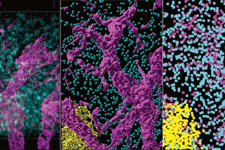
Improvement of Imaging Techniques to Understand Organelle Membrane Cell Dynamics
Understanding cell functions in normal and tumorous tissue is a key factor in advancing research of potential treatment strategies and understanding why some treatments might fail. Single-cell…
Loading...

Image Gallery: THUNDER Imager
To help you answer important scientific questions, THUNDER Imagers eliminate the out-of-focus blur that clouds the view of thick samples when using camera-based fluorescence microscopes. They achieve…
Loading...
From Organs to Tissues to Cells: Analyzing 3D Specimens with Widefield Microscopy
Obtaining high-quality data and images from thick 3D samples is challenging using traditional widefield microscopy because of the contribution of out-of-focus light. In this webinar, Falco Krüger…
Loading...

Studying Human Brain Development and Disease
Neural spheroids created from human induced pluripotent stem cells (iPSCs) provide effective and novel tools for studying brain development, as well as the underlying pathological mechanisms of…
Loading...

An Introduction to Computational Clearing
Many software packages include background subtraction algorithms to enhance the contrast of features in the image by reducing background noise. The most common methods used to remove background noise…
Loading...
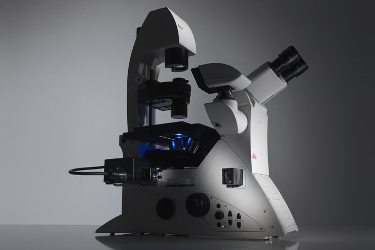
Factors to Consider When Selecting a Research Microscope
An optical microscope is often one of the central devices in a life-science research lab. It can be used for various applications which shed light on many scientific questions. Thereby the…
Loading...
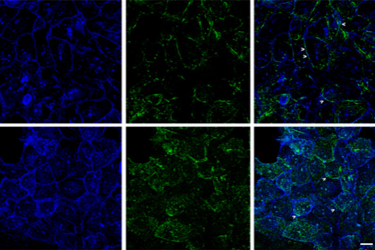
 - THUNDER Imager 3D Cell Culture Influenca virus – red, cilia – green, Nuclei – blue.")
How Can Immunofluorescence Aid Virology Research?
Modern virology research has become as crucial now as ever before due to the global COVID-19 pandemic. There are many powerful technologies and assays that virologists can apply to their research into…