
Mouse lymphnode - Getting the complete picture for inflammation research
A part of the work at the Keppler lab involves the use of pre-clinical models for inflammatory disease to understand the interplay between cells during inflammation. In order to understand the distribution of the cells within the organ, the lab researchers needed to change from imaging 2D cryo-sections of lymph nodes to a method that is capable of viewing the whole organ.
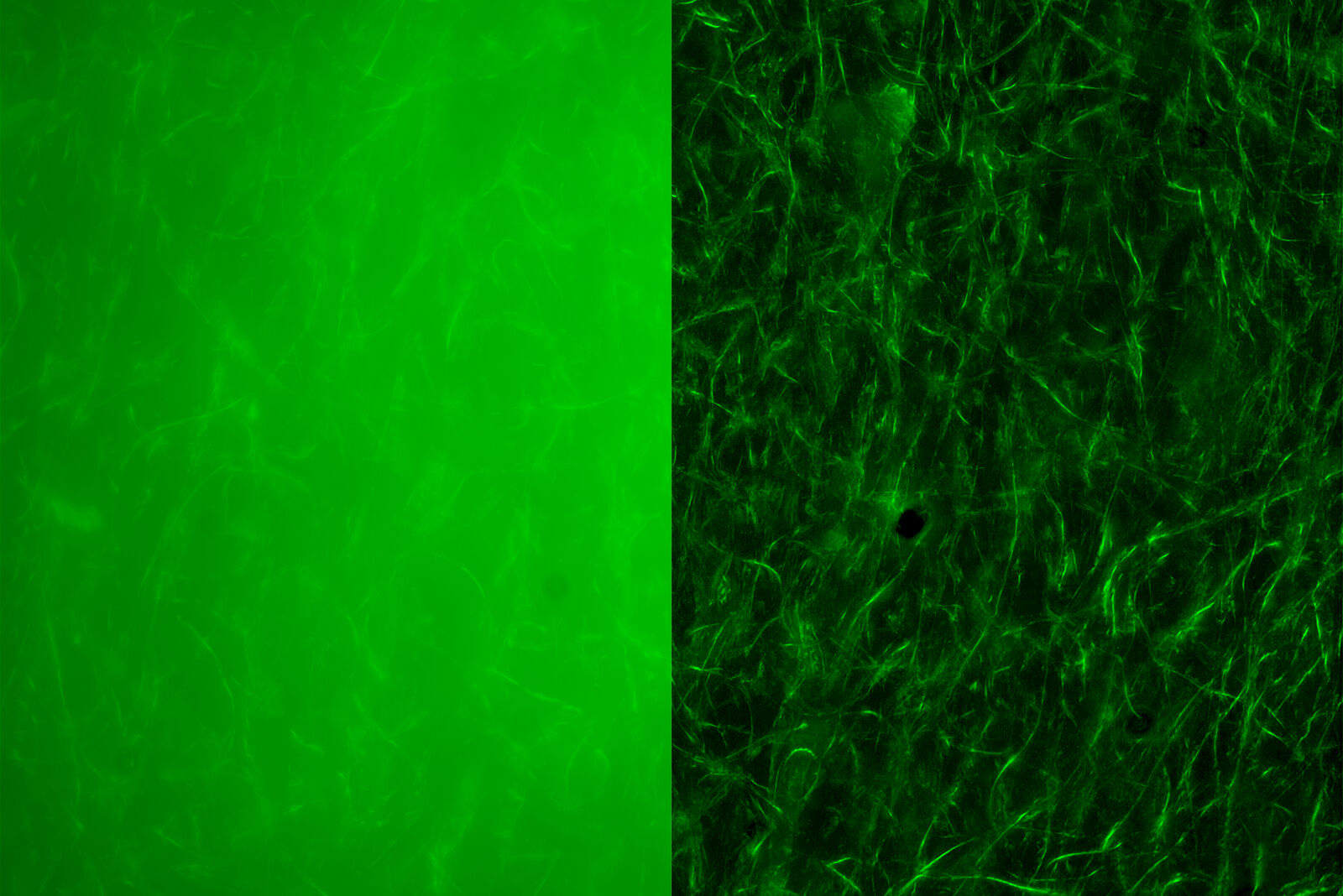
They developed a clearing protocol to prepare the lymph nodes for imaging. Initially, they performed the imaging experiments using a confocal microscope, but this way took up to 18 hours to image just half of a lymph node. They could not use traditional widefield systems, because the autofluorescence coming from the tissue makes it impossible to see any significant amount of the desired signal.
Only with a THUNDER Imager, we could consider using a widefield-based system to study our organs
Dr. Selina Keppler, Munich, Germany.
Image the cells of interest within the lymph node in only a few minutes
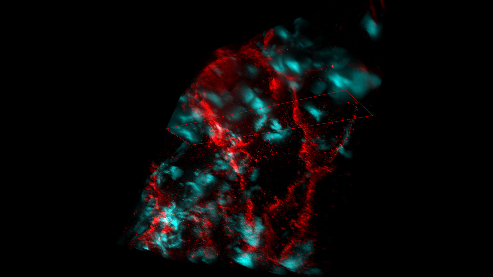
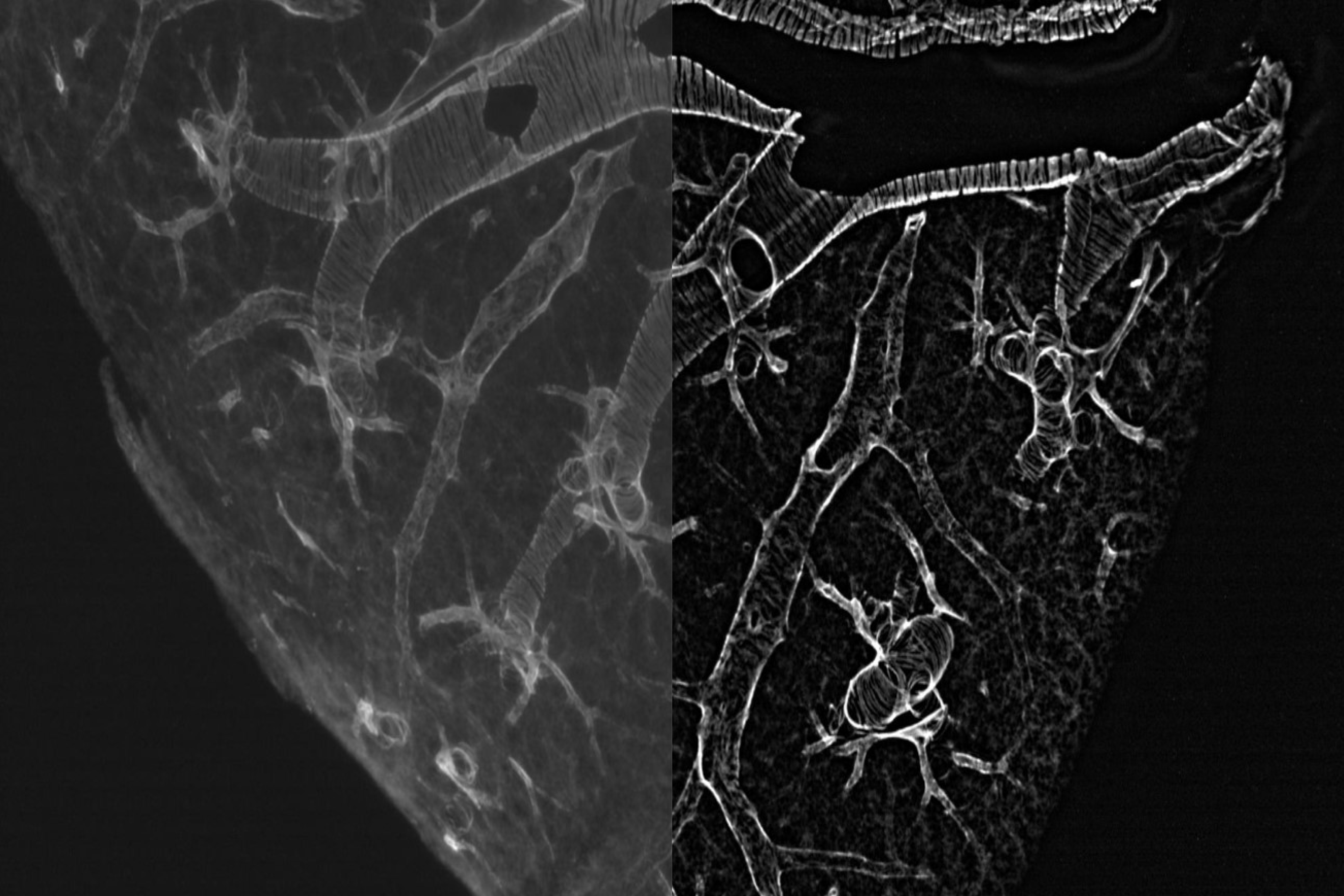
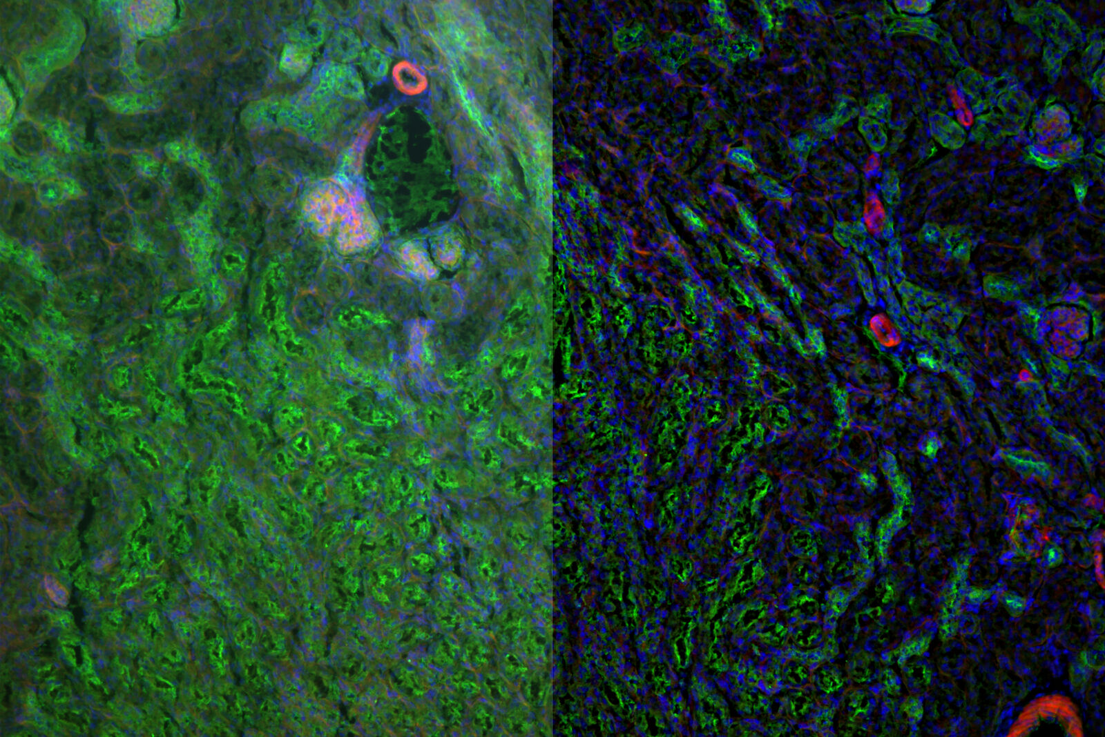
Using a THUNDER Imager, the Keppler lab researchers found that they could clearly image the cells of interest within the lymph node in only a few minutes. In the example shown, they were able to clearly study the distribution of follicular cells (yellow), and blood vessels (magenta). Then they went on to study differences in wild type vs. inflamed lymph nodes. Having this initial overview in just 15 minutes helped them develop a more efficient screening workflow where they could then select the areas they wanted to analyze in detail using confocal microscopy enabling more detail segmentation or single cell analysis.
The THUNDER Imager has made our research more efficient, in terms of time savings, and more complete, as we can now look at whole structures and are not limited at just looking at parts of the organ
Dr. Selina Keppler, Munich, Germany.
Arabidopsis thaliana root - THUNDER Imager
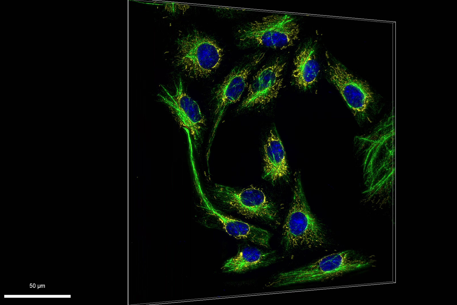
Hela Cells - THUNDER Imager
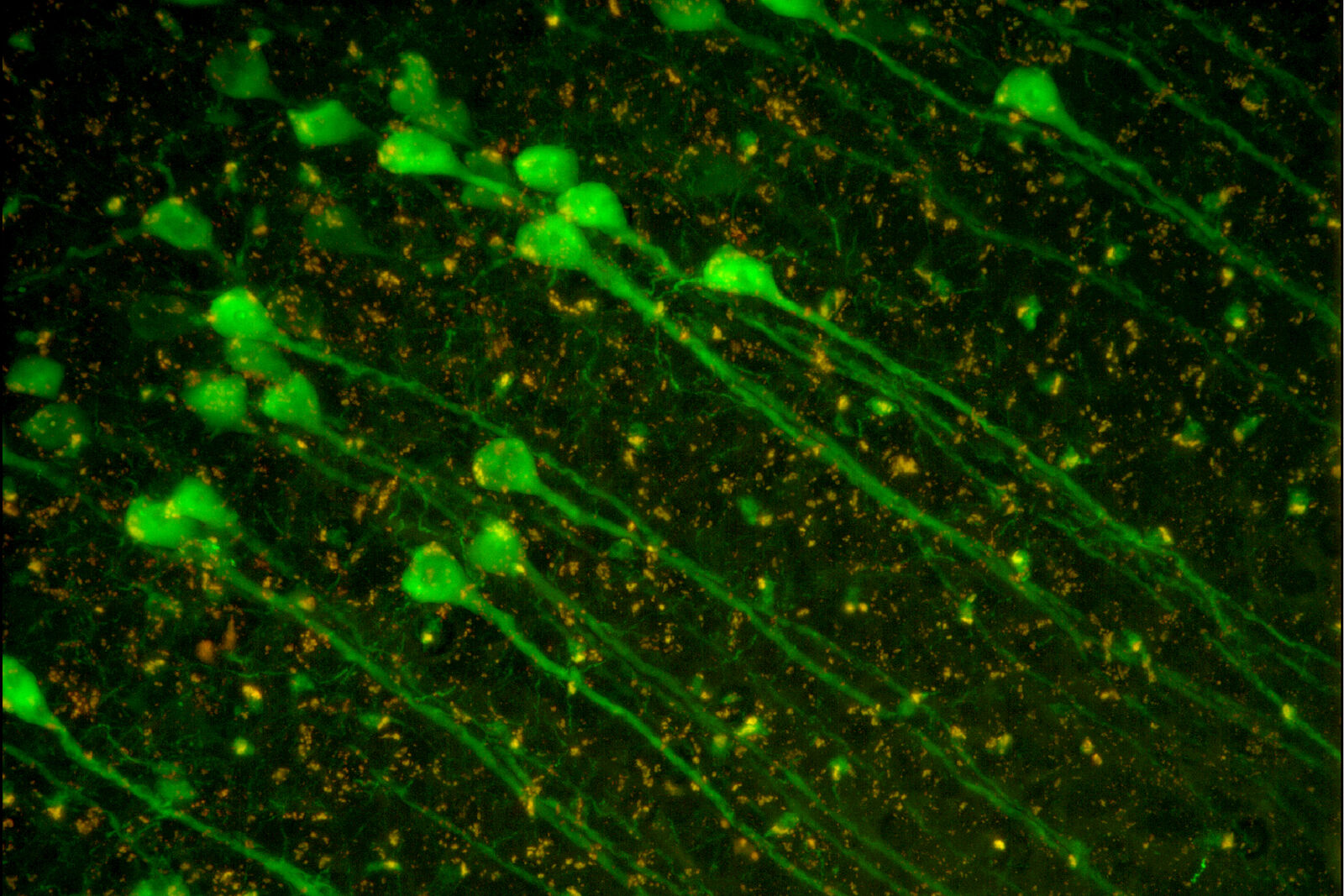
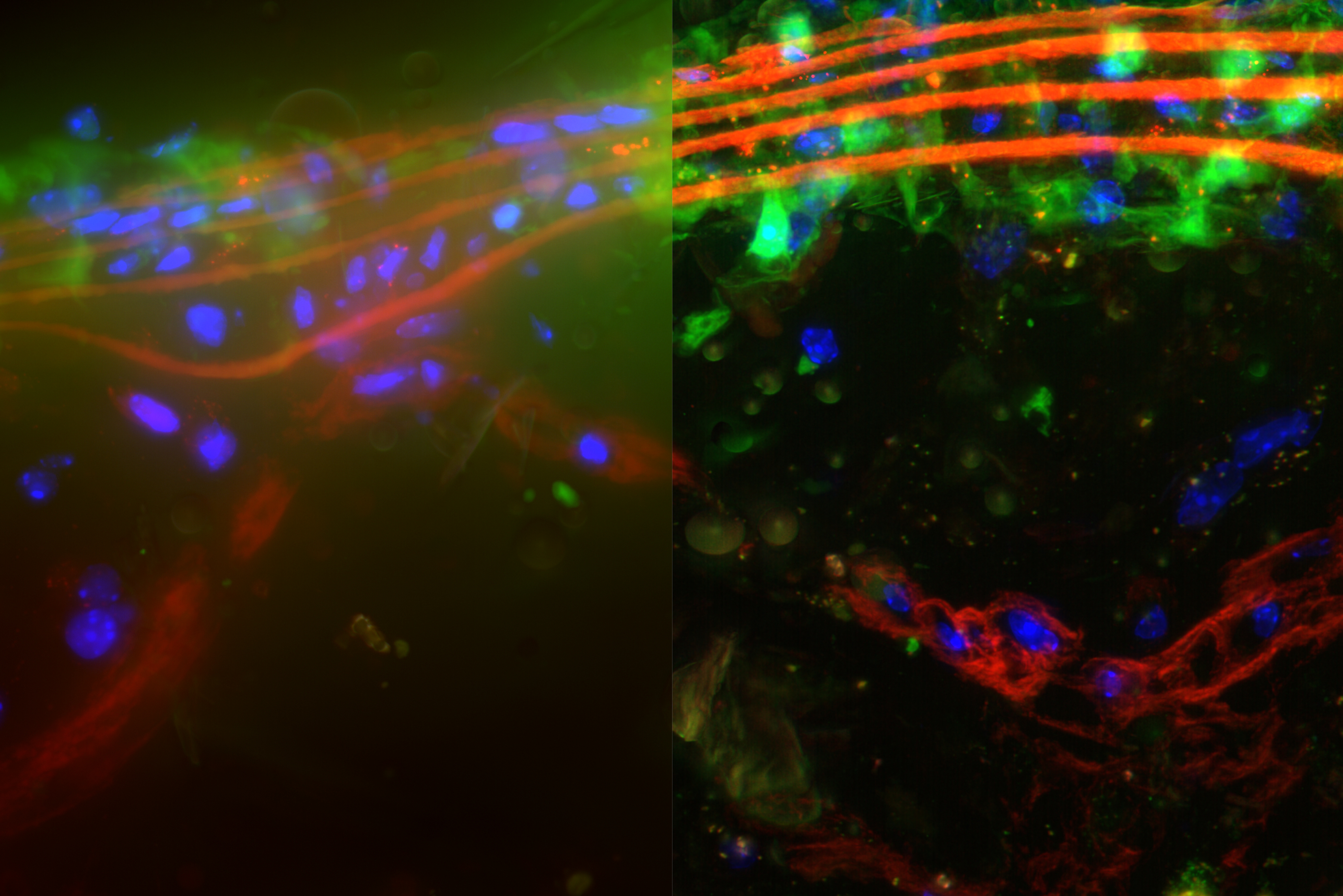
Mouse brain cortex - THUNDER Imager

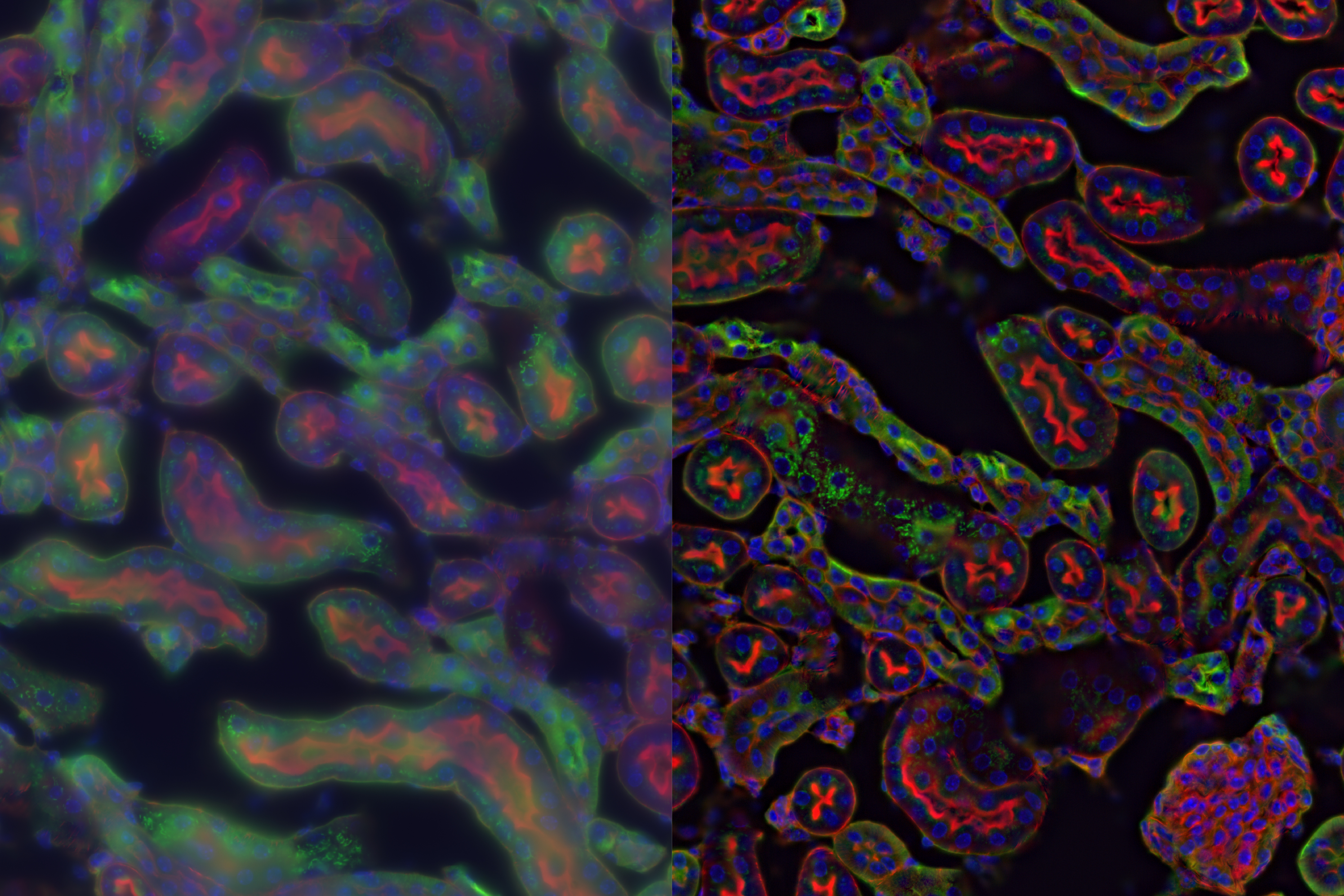
Mouse embryonic kidney - THUNDER Imager

Murine esophageal organoids - THUNDER Imager

Sample courtesy of Dr. Fabio Tadeu Arroso Martins, Tampere University, Finland. Sample imaged by Janne Ylärinne, PhD, Immuno Diagnostic Oy.
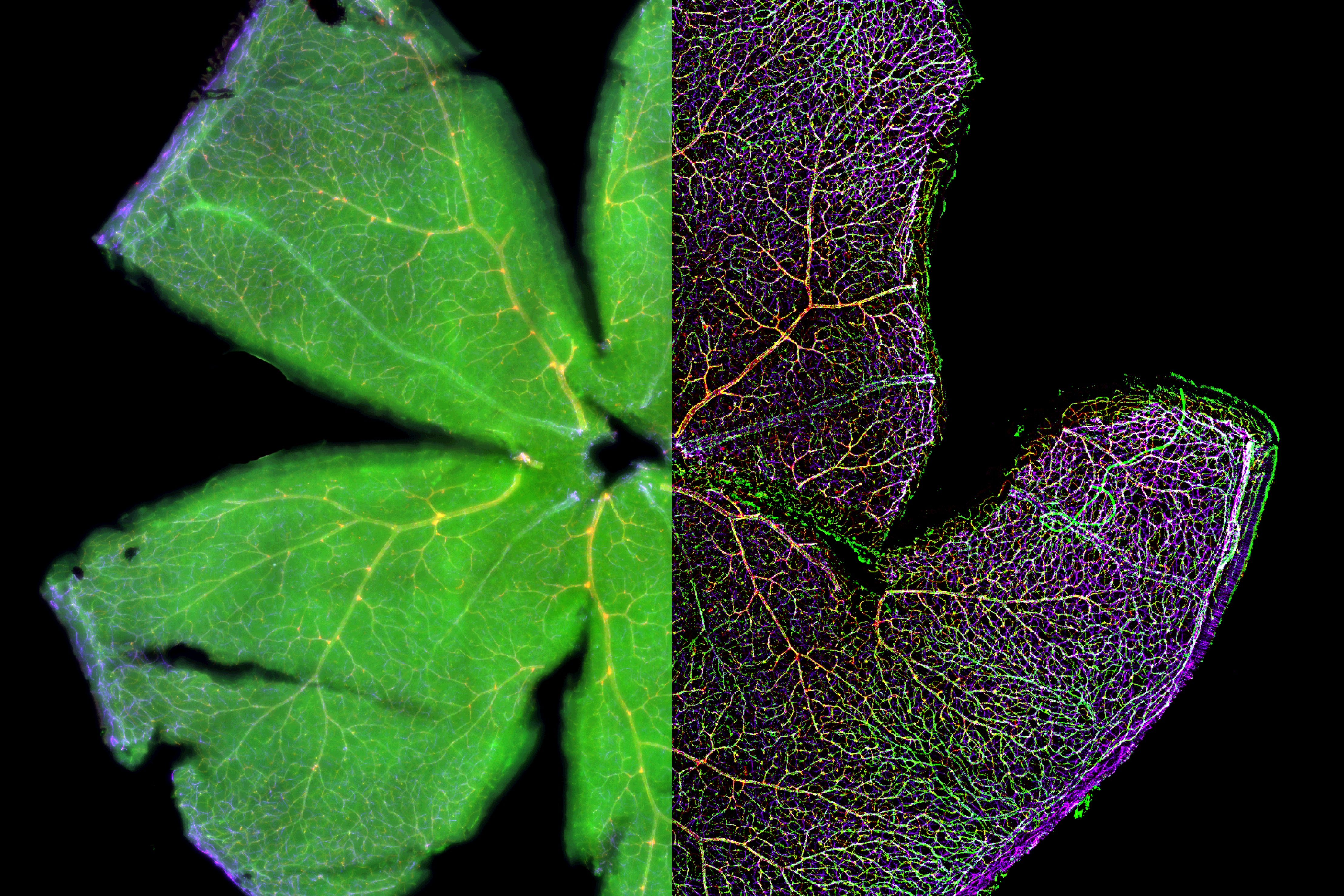
Mouse whole mount retina - THUNDER Imager

1 mm mouse brain section - THUNDER Imager
C. elegans. Transgenic GFP - THUNDER Imager
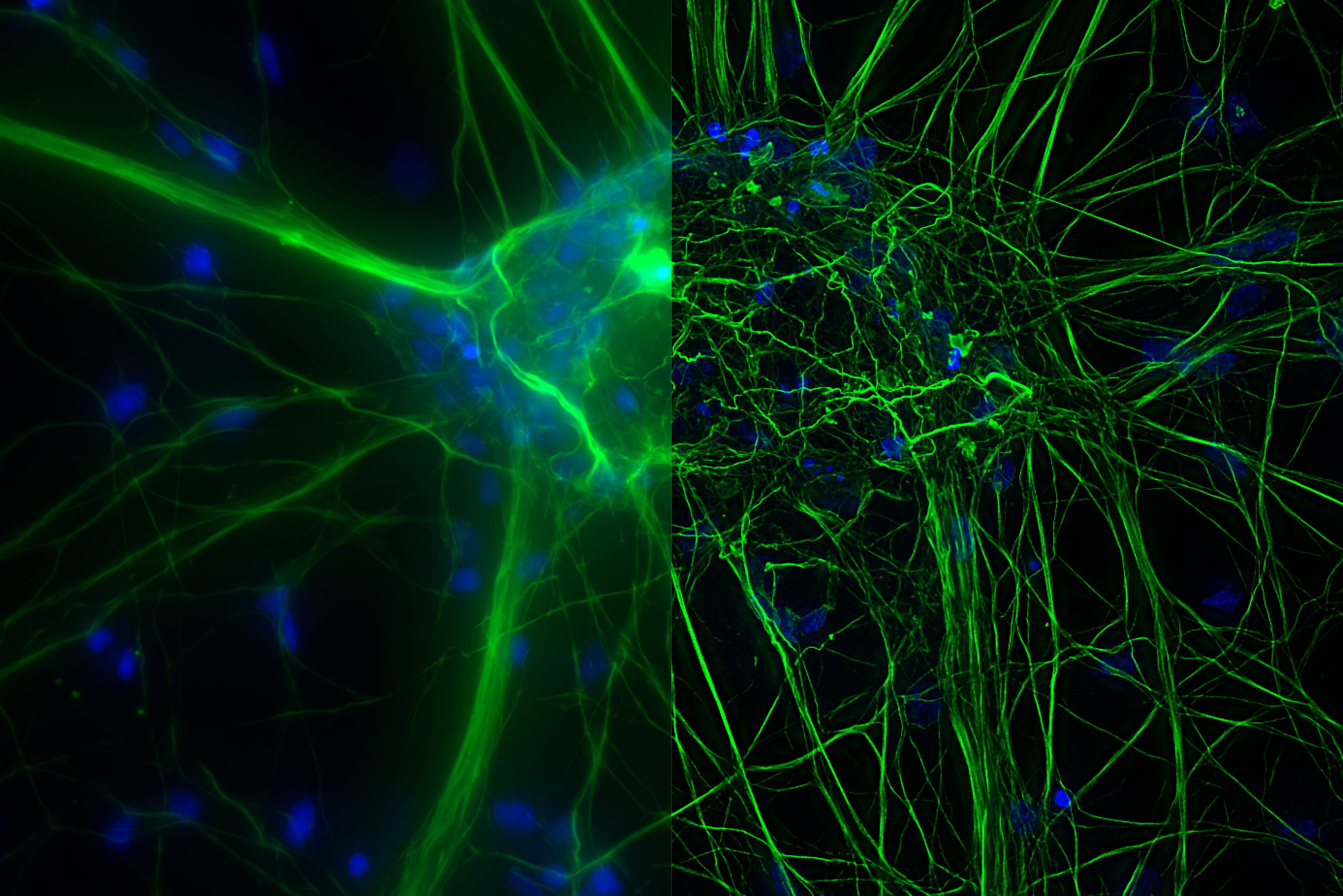
Mouse cortical neurons - THUNDER Imager
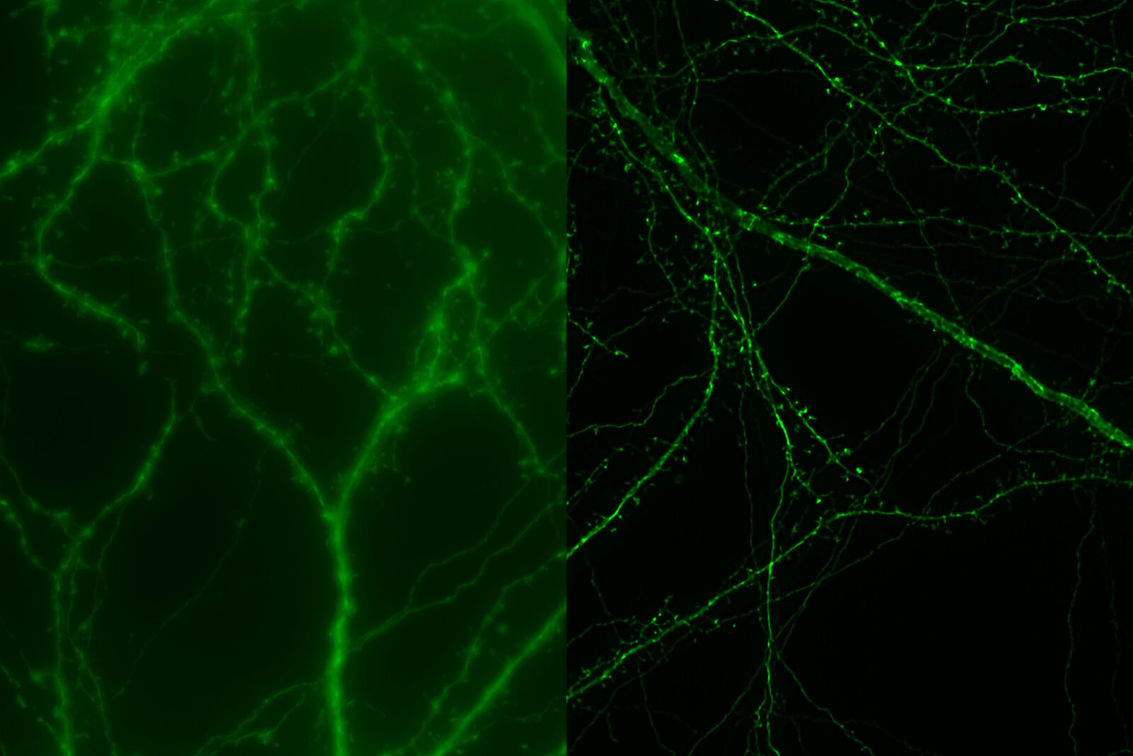
Image courtesy of Prof. Hui Guo, School of Life Sciences, Central South University, China
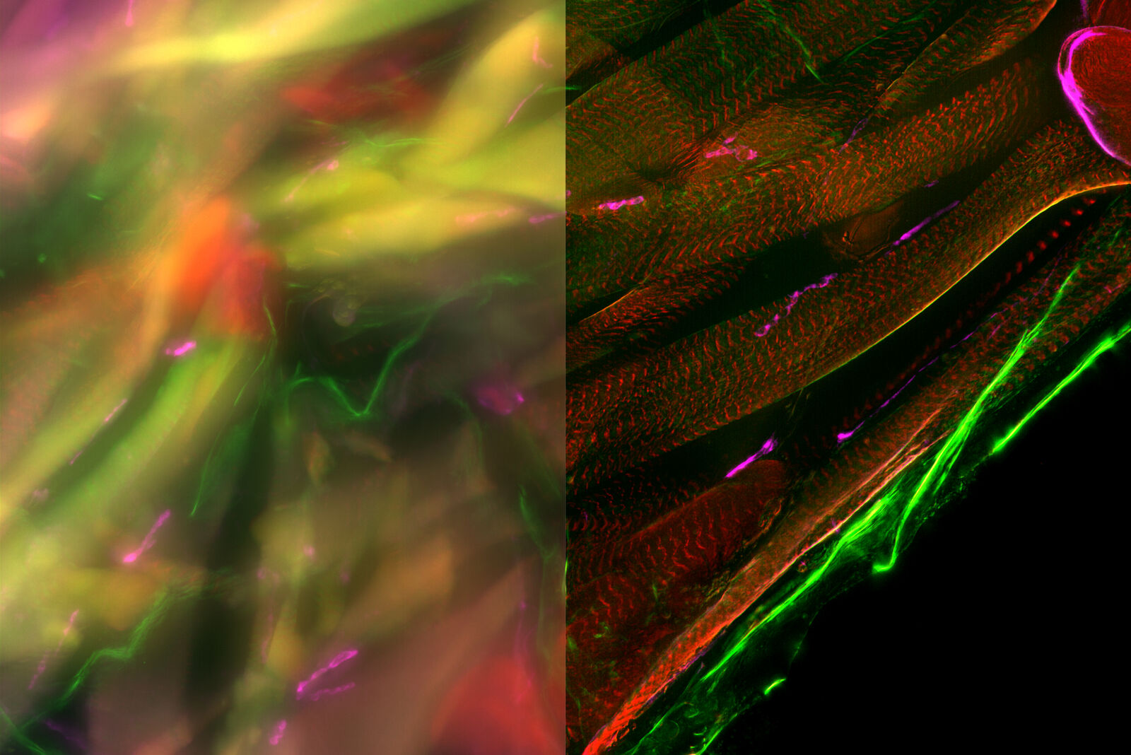
Adult Drosophila muscle - THUNDER Imager
Drosophila Embryo - THUNDER Imager
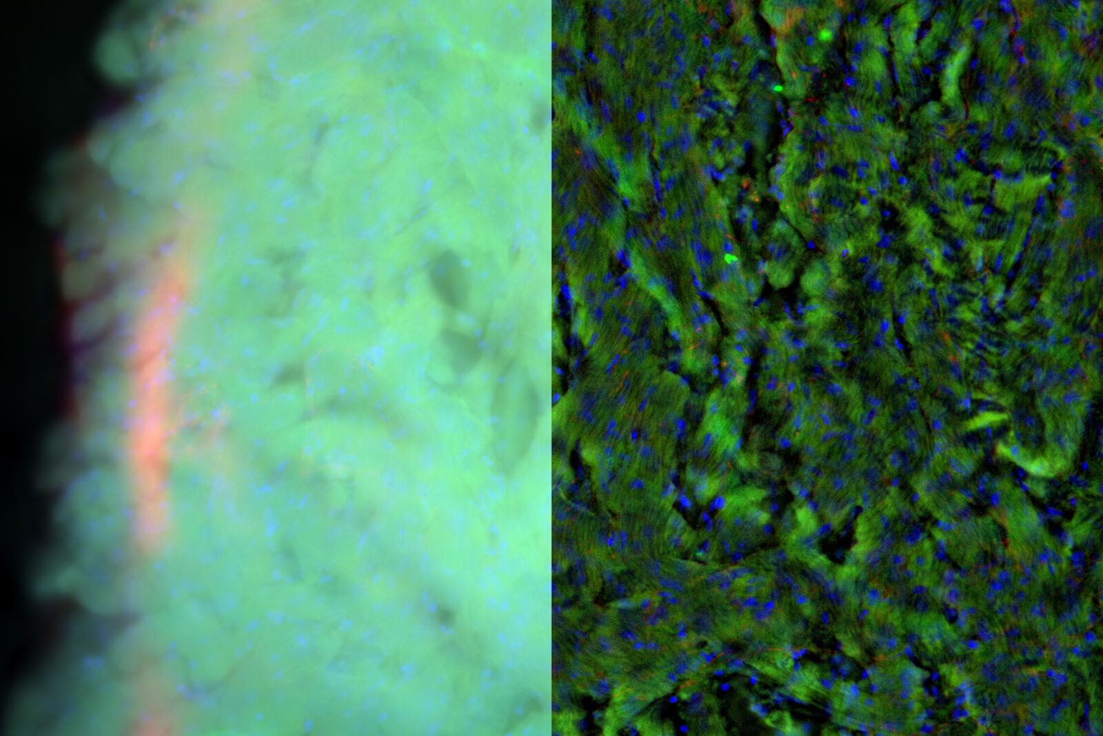
Mouse heart - THUNDER Imager 3D Cell Culture
Drosophila Follicles - THUNDER Imager 3D Cell Culture
Cortical neurons - THUNDER Imager

Image courtesy of Wei Wang, USA
Microglia in hippocampal region - THUNDER Imager
Organoid Cluster - THUNDER Imager
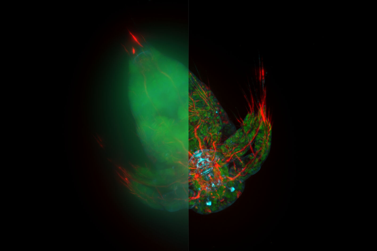
Cranial nerves development - THUNDER Imager 3D Cell Culture
Pancreatic islet - THUNDER Imager 3D Cell Culture
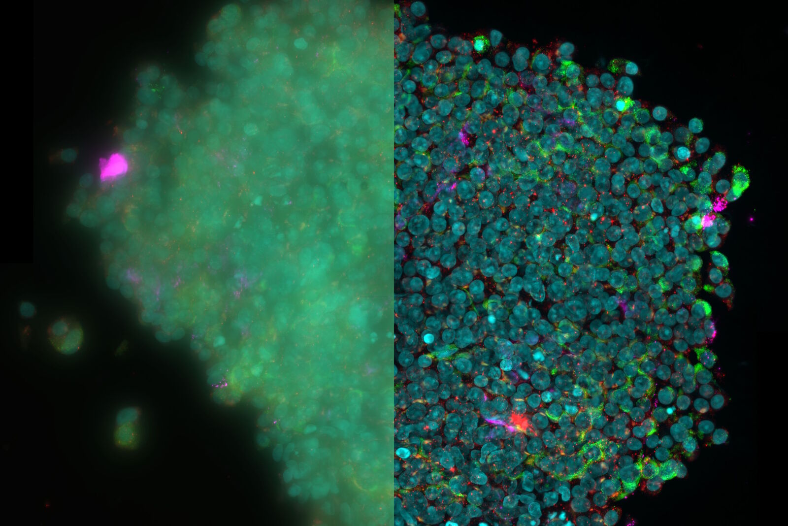
Image courtesy of the Matthias Von Herrath Lab at the La Jolla Institute of Immunology, La Jolla, CA., USA
Mouse Brain - THUNDER Imager

Image courtesy of Maheedhar Kodali, Ashok K. Shetty, Texas A&M University, USA
Pollen Flower - THUNDER Imager
C. elegans Gonades - THUNDER Imager
Mouse aorta - THUNDER Imager 3D Live Cell
Mouse aorta - THUNDER Imager 3D Live Cell
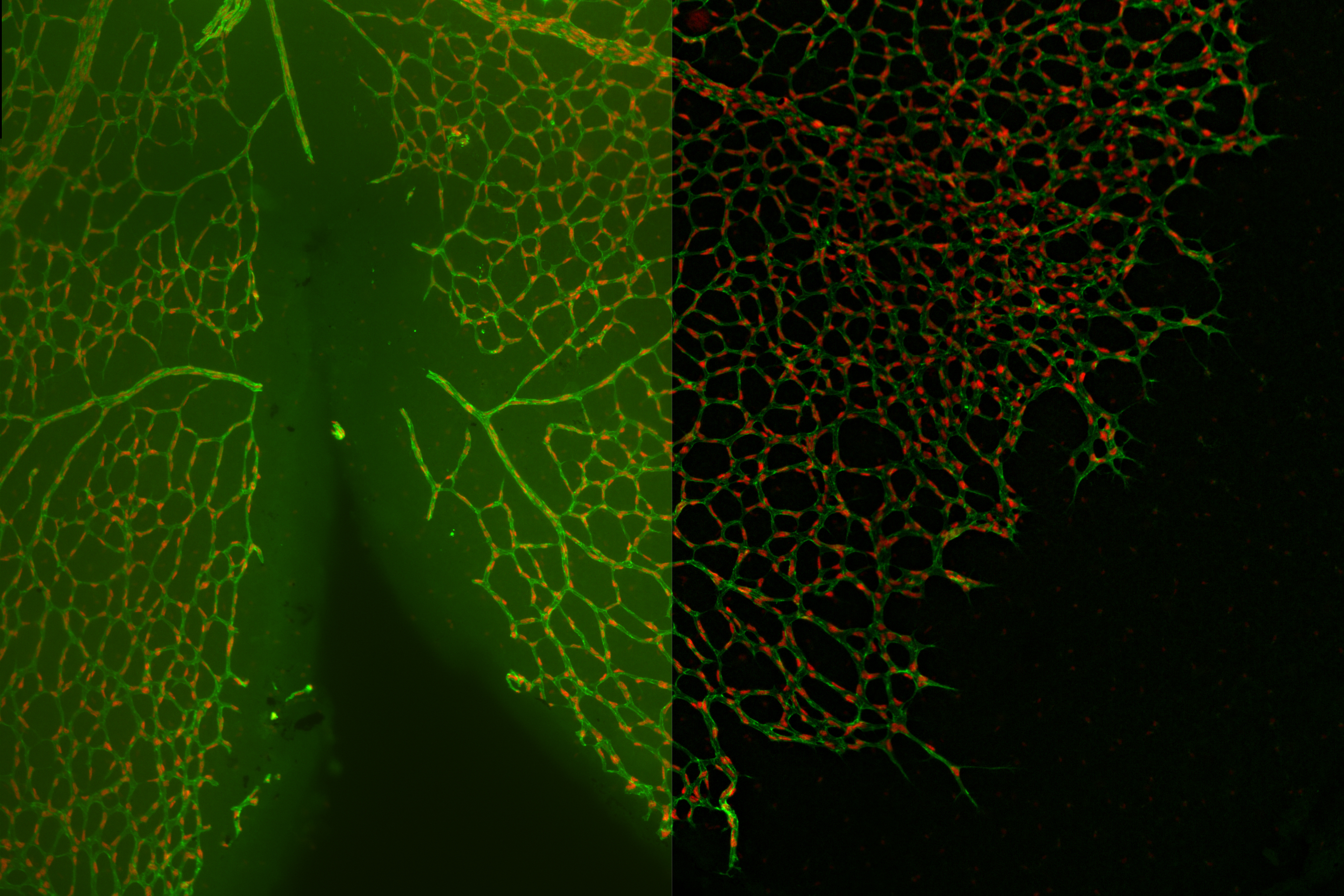
Mouse Dystrophin staining on muscle fibres - THUNDER Imager
Infected MDCK epithelial cells - THUNDER Imager

Objective: HC PL APO 40x/0.95 DRY.
Image courtesy of Dr. Mark Jepson, Bristol University, Bristol, UK.
Ferret brain, rabies infected - THUNDER Imager 3D Cell Culture

Image courtesy of Dr. Stefan Finke, Friedrich-Loeffler- Institute, Riems, Germany.
Influenza in lung epithelial cells (porcine) - THUNDER Imager 3D Cell Culture
Lung Organoid - THUNDER Imager 3D Cell Culture
Pseudoislets (pancreatic beta cells) - THUNDER Imager 3D Cell Culture
Locust ganglion - THUNDER Imager 3D Tissue
HeLa cell spheroid - THUNDER Imager 3D Live Cell & 3D Cell Culture
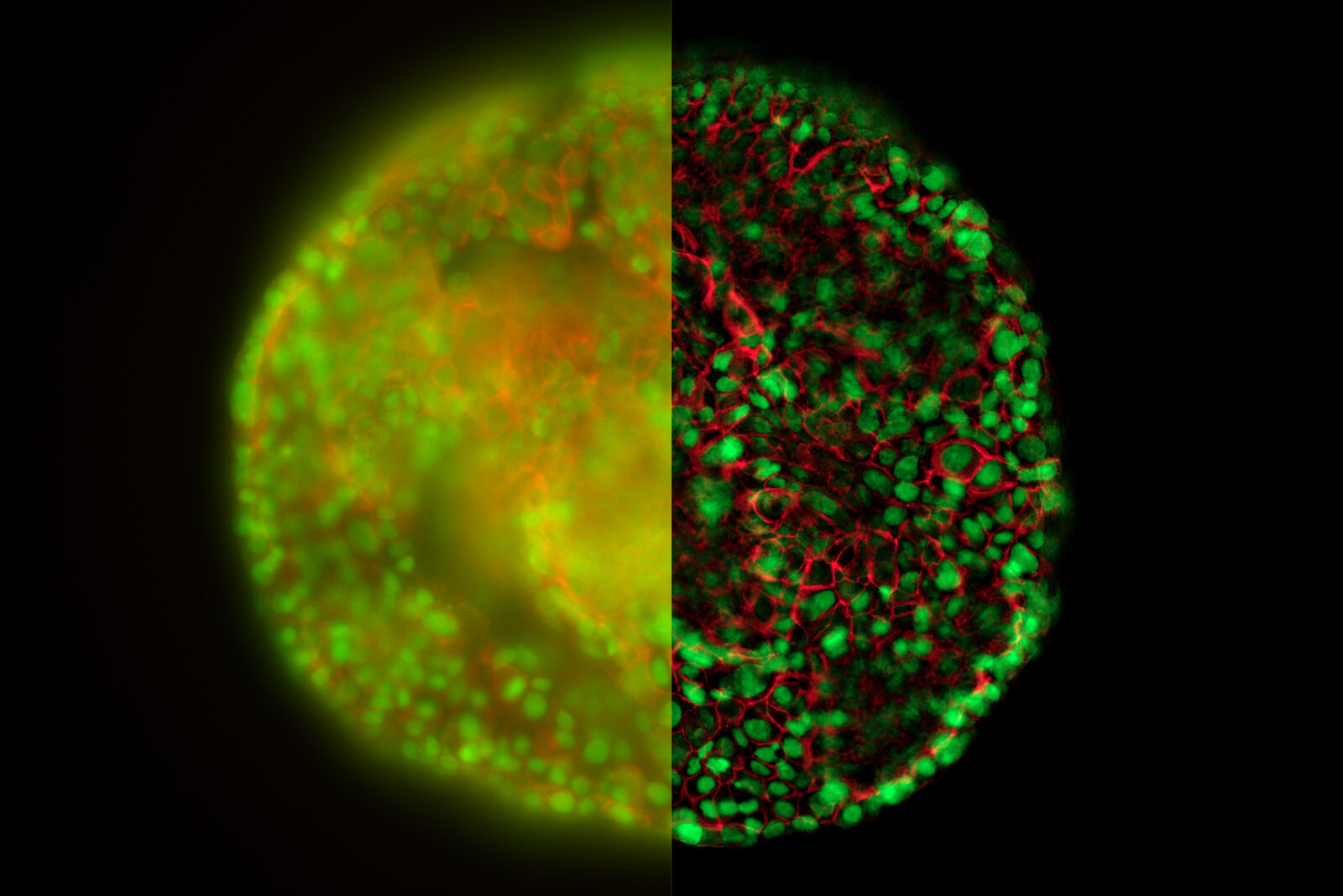
Image taken by Dr. Jan Schumacher, Leica Microsystems, Germany.
Zebrafish - THUNDER Imager 3D Live Cell & 3D Cell Culture
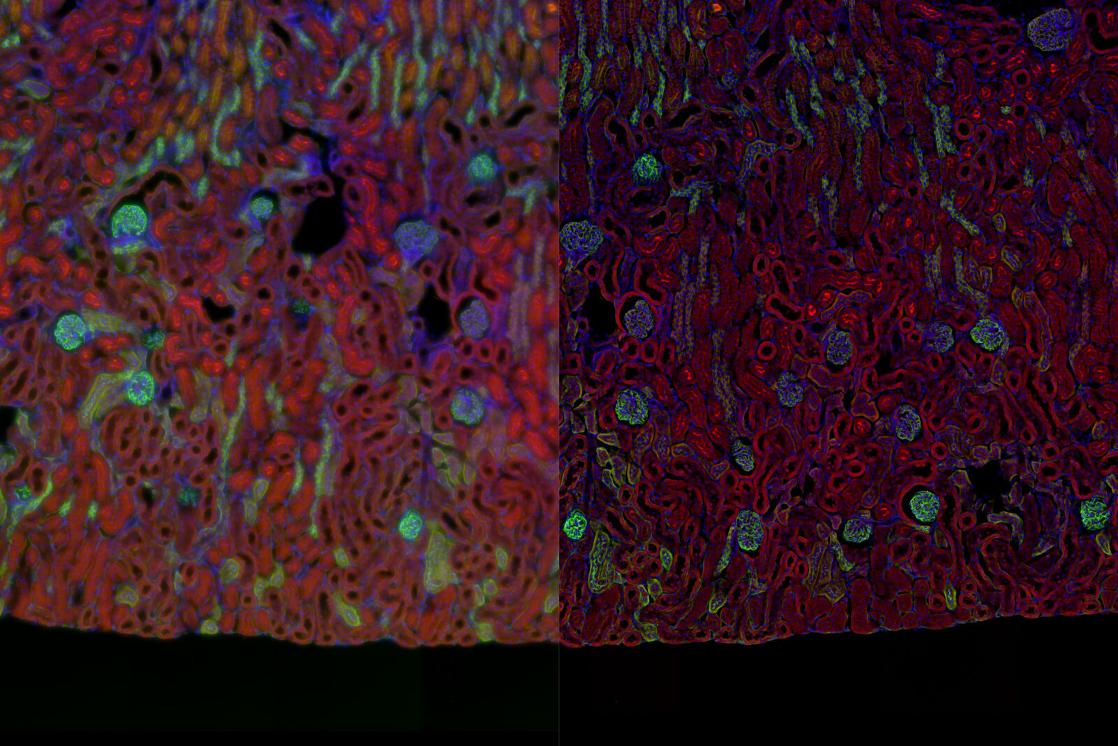
Mouse kidney section - THUNDER Imager Tissue 3D
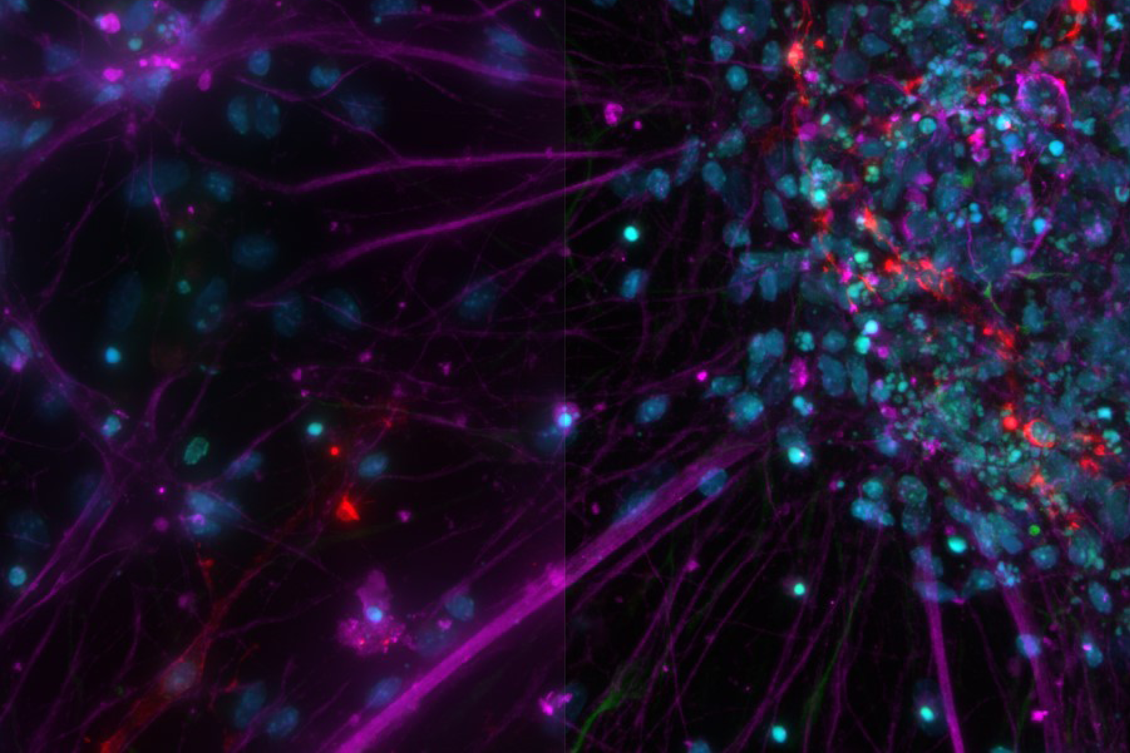
Cultured Cortical Neurons - THUNDER Imager 3D Cell Culture
Cultured VERO cells - THUNDER Imager 3D Cell Culture
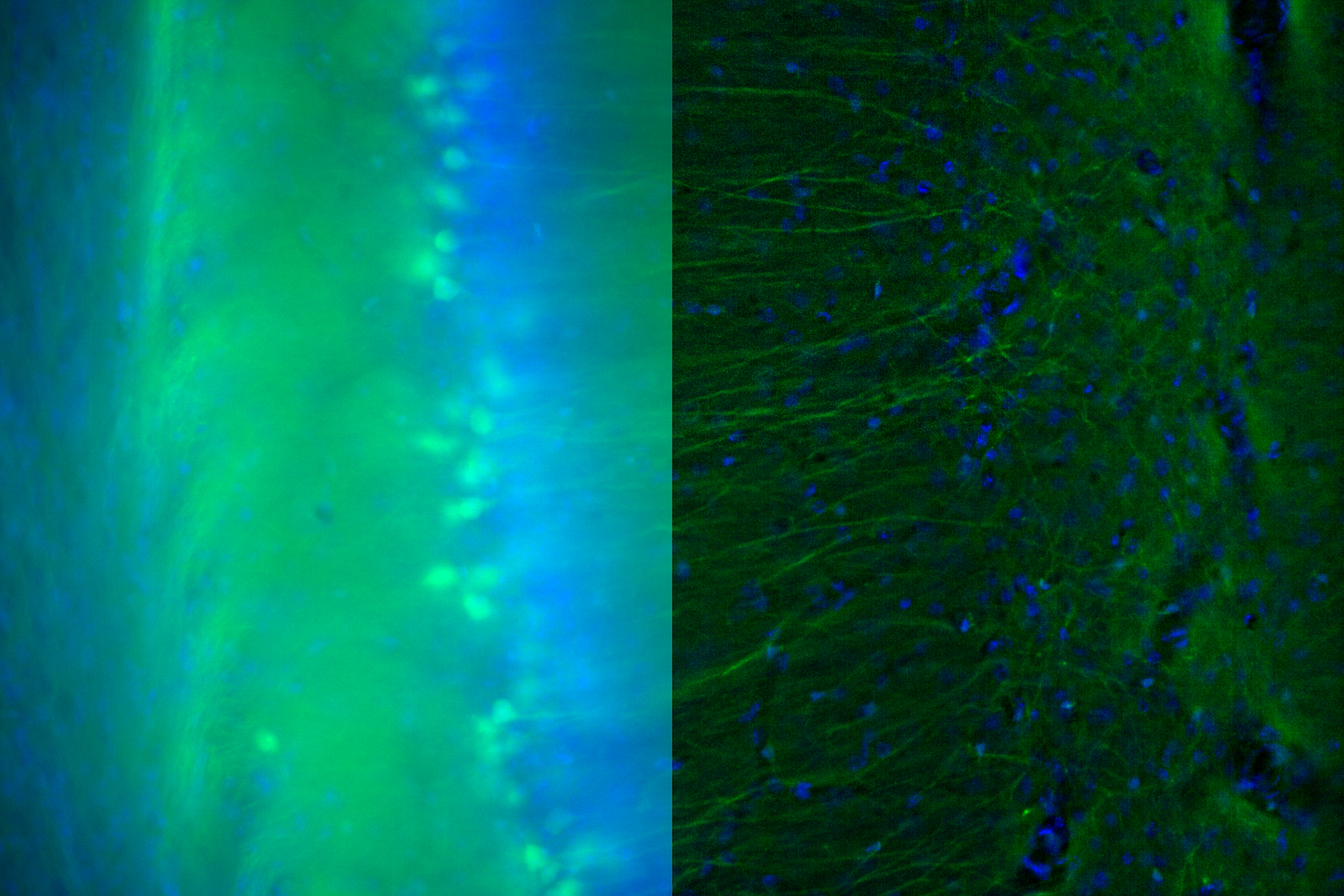
YFP mouse brain slice - THUNDER Imager Tissue
Drosophila third instar larval - THUNDER Imager Tissue
Cyclops - THUNDER Imager Tissue
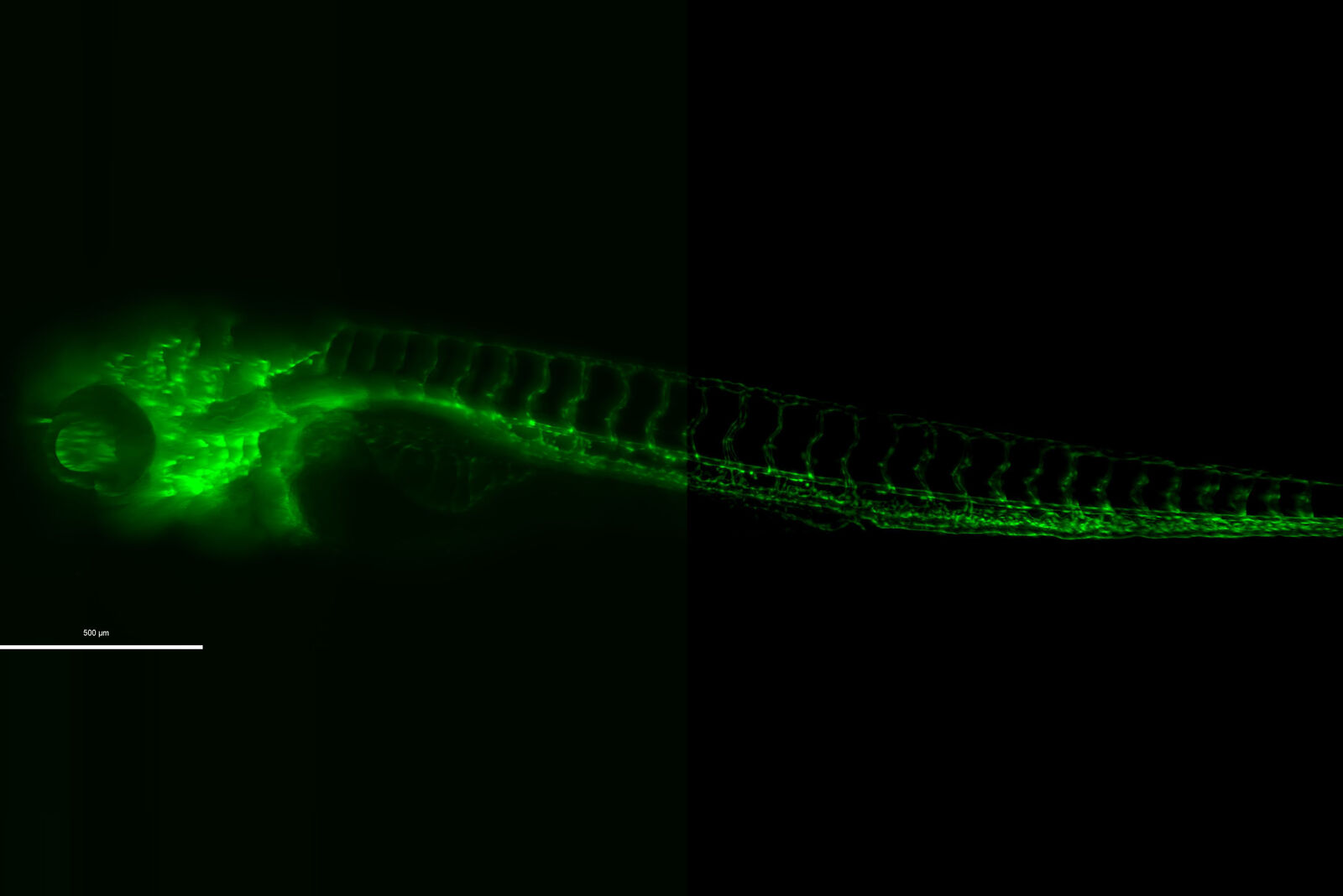
Zebrafish – THUNDER Imager Model Organism
Mouse - THUNDER Imager Model Organism
Organoid - THUNDER Imager Model Organism
Drosophila brain - THUNDER Imager 3D Live Cell
Honeybee leg - THUNDER Imager Model Organism
Paramecium - THUNDER Imager
Mouse kidney section - THUNDER Imager 3D Tissue
Mouse kidney section - THUNDER Imager 3D Tissue
Smooth Muscle - THUNDER Imager Model Organism
Courtesy of Dr. Mario Boehm, University of Giessen, Germany
Primary culture, rat - THUNDER Imager 3D Cell Culture
Mouse kidney section - THUNDER Imager 3D Tissue
Zebrafish heart - THUNDER Imager Tissue
Zebrafish embryo – THUNDER Imager Model Organism
C2C12 cells - THUNDER Imager 3D Live Cell & 3D Cell Culture
Images courtesy of Dr. Lucas Smith, Department of Neurobiology, Physiology and Behavior, College of Biological Sciences, University of California at Davis, Davis CA, USA.
Virally labeled neuron - THUNDER Imager Model Organism
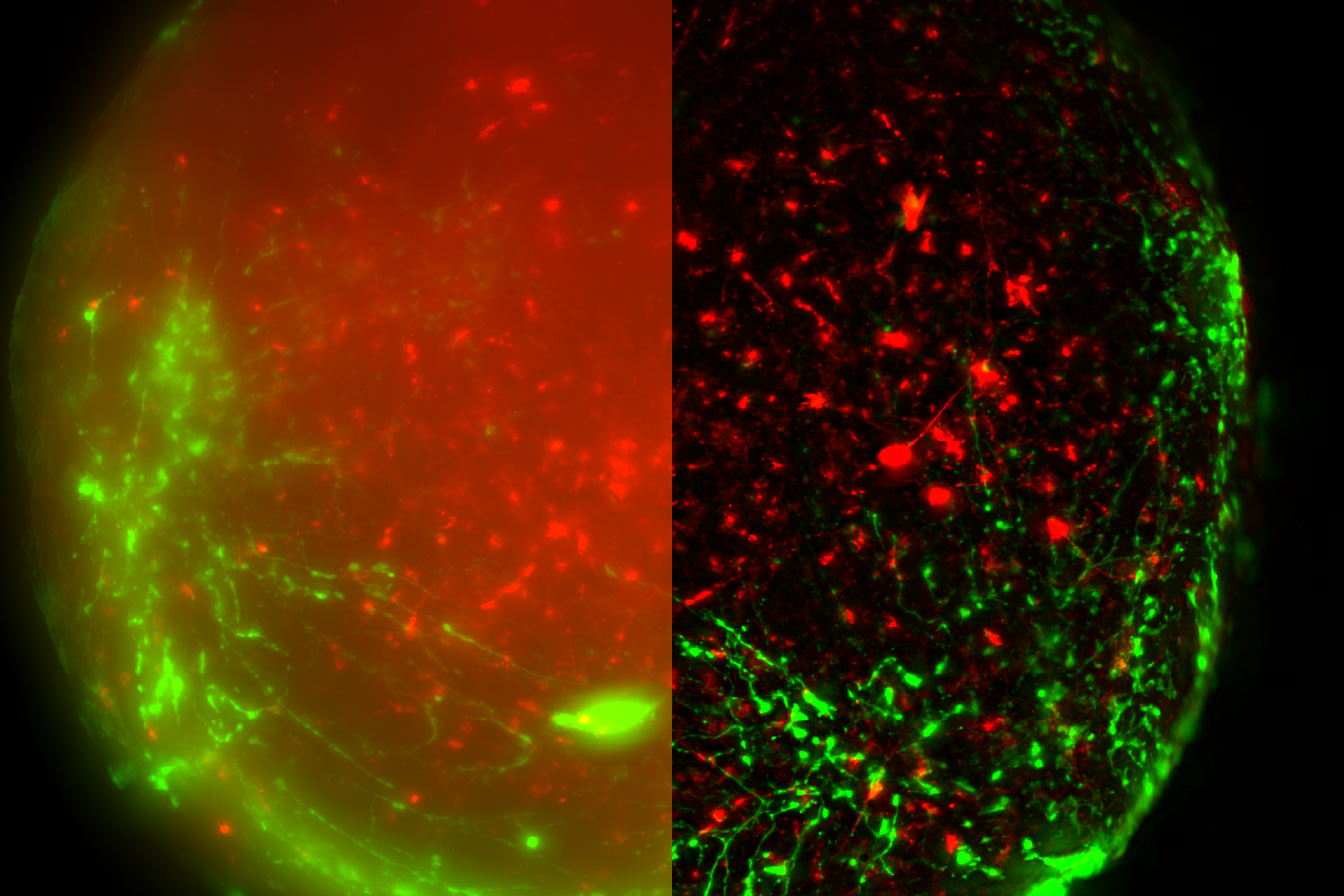
Images courtesy of Dr. Fikri Birey from the Dr. Sergiu Pasca laboratory, CA, USA.
Transgenic Drosophila photoreceptors of the eye - THUNDER Imager 3D Cell Culture
Cochlea cell - THUNDER Imager 3D Tissue
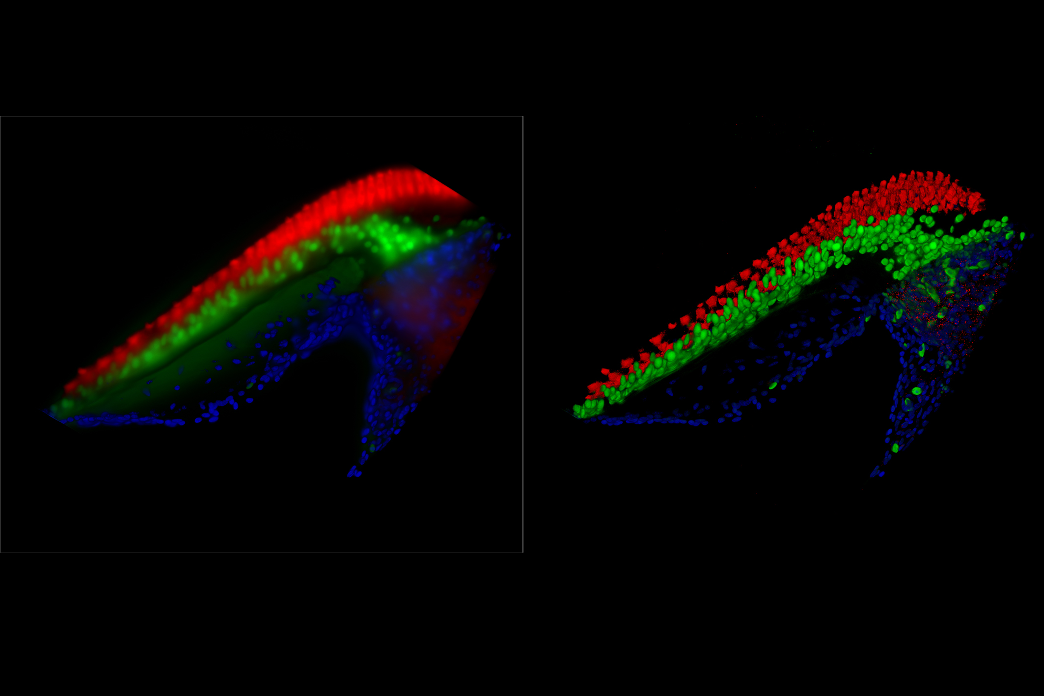
Image courtesy of Amanda Janesick, CA, USA.
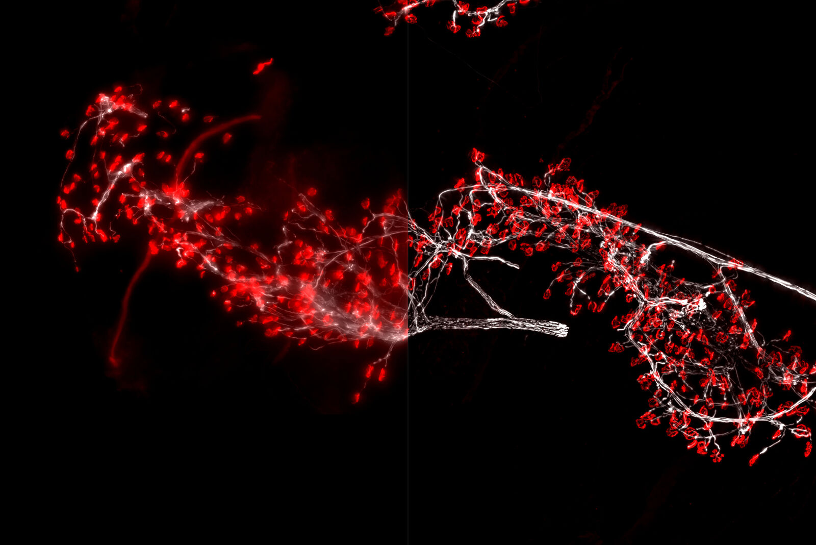
Neuromuscular junctions - THUNDER Imager 3D Tissue
Sea anemone polyp (Exaiptasia pallida) - THUNDER Imager Model Organism
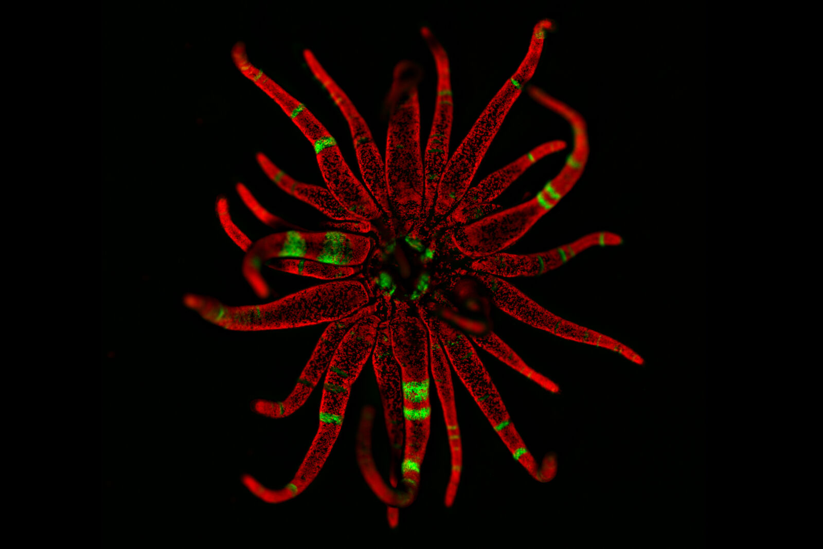
Image courtesy of Christian Renicke, CA, USA.
Mouse lung - THUNDER Imager Tissue
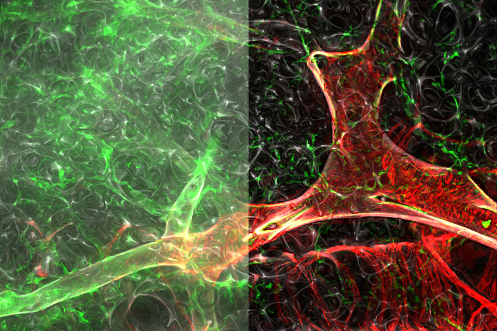
Image courtesy of Ross Metzger, CA, USA.
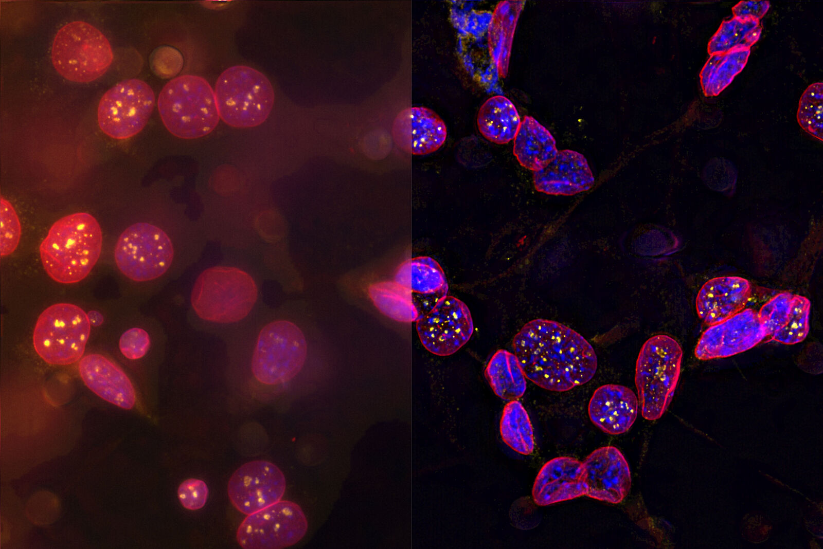
Hippocampus showing an amyloid plaque - THUNDER Imager 3D Cell Culture
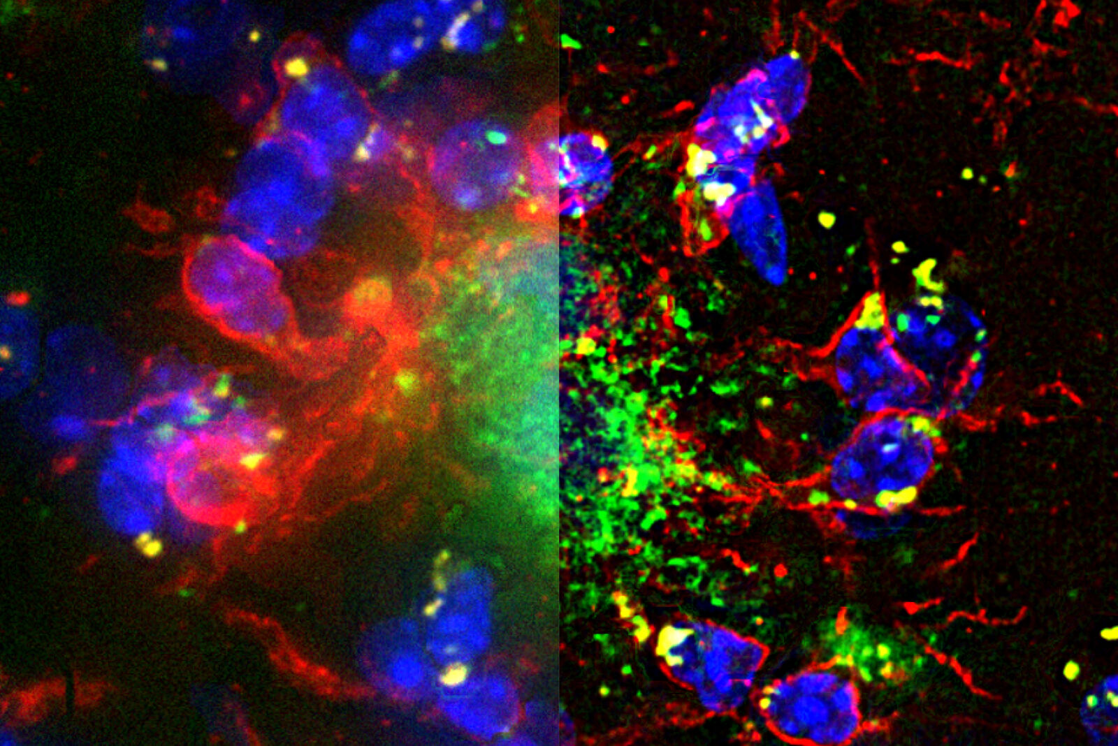
(green, stained with 6E10 antibody, marker of anti-beta amyloid) surrounded by microglia/microphages (red, stained with Anti-Abi1 antibody; blue, DAPI).
Left – Raw data with Extended Depth of Field projection;
Right – Large Volume Computationally Cleared Z-stack with Extended Depth of Field projection.
Images courtesy of Prof. Mehrdad Shamloo, CA, USA.
Neural crest (NC) embryonic cell population - THUNDER Imager 3D Cell Culture
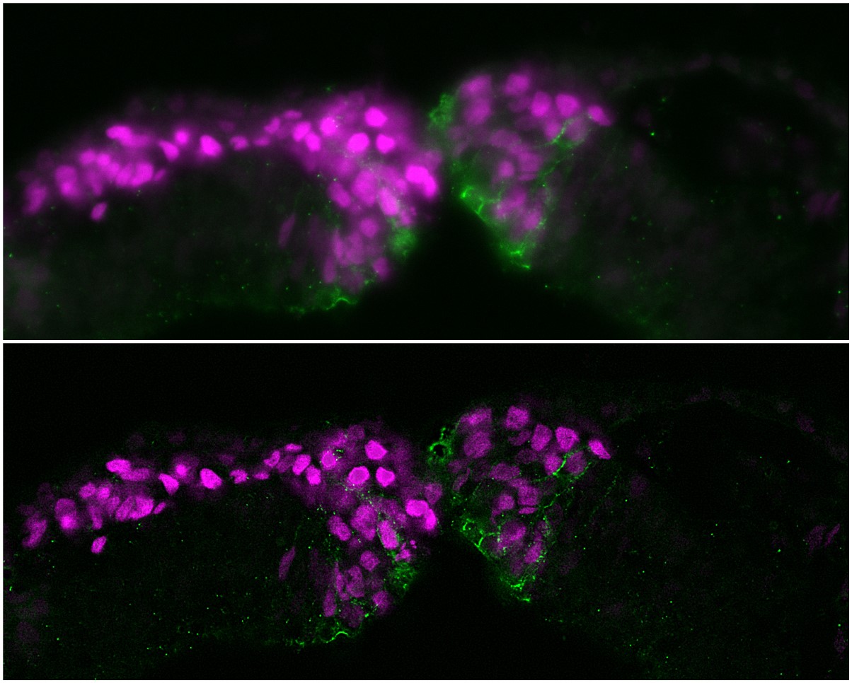
Image courtesy of Michael Piacentino, BronnerLab, California Institute of Technology, Pasadena, CA, USA.
Adult mouse ovary - THUNDER Imager 3D Cell Culture
C. elegans - THUNDER Imager Model Organism

The green punctae (GFP) are APs while the ALs quench GFP in the acidic environment and emit only the mCherry signal.
Image courtesy of Dr. Aditi U. Gurkar, University of Pittsburgh, PA, USA
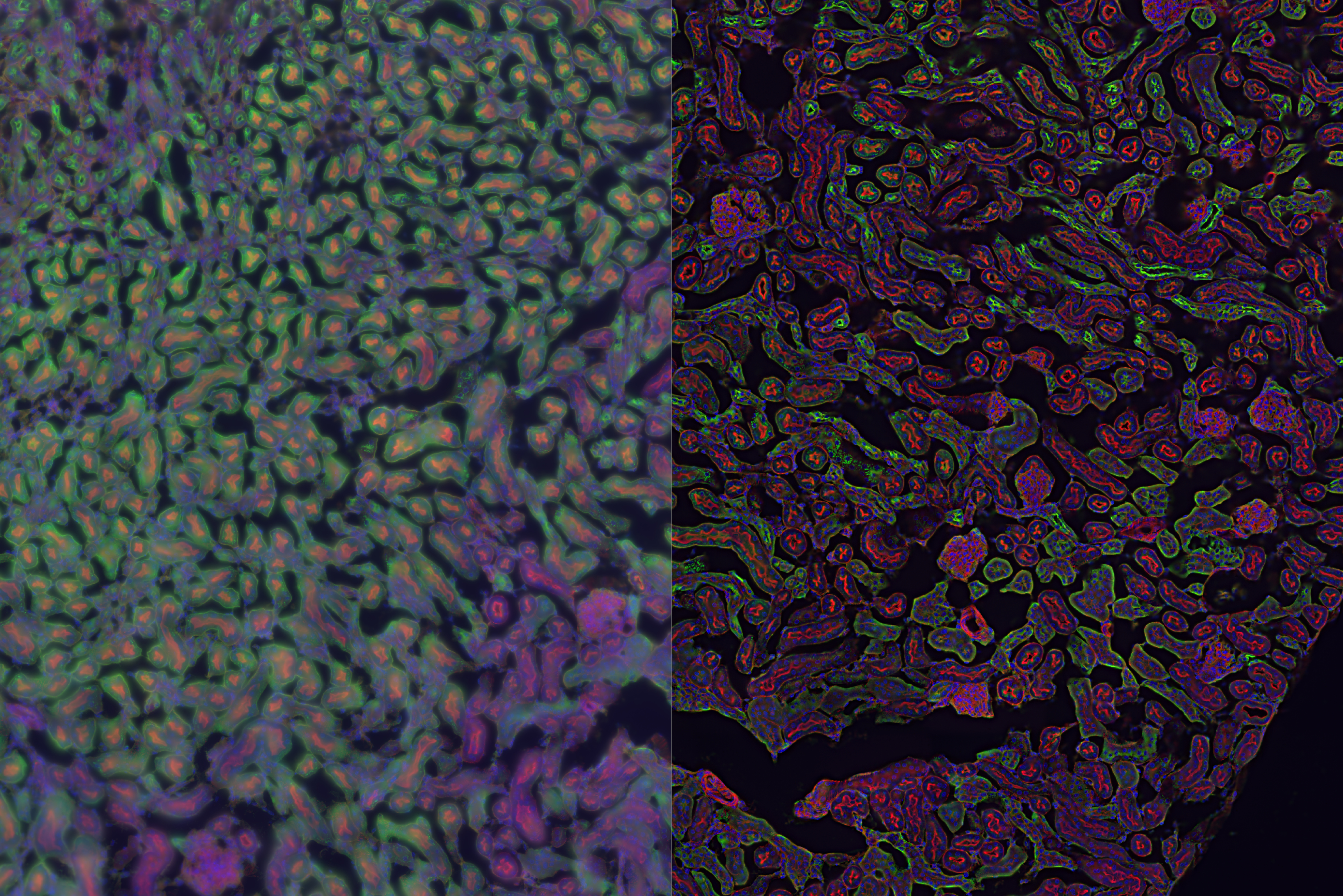
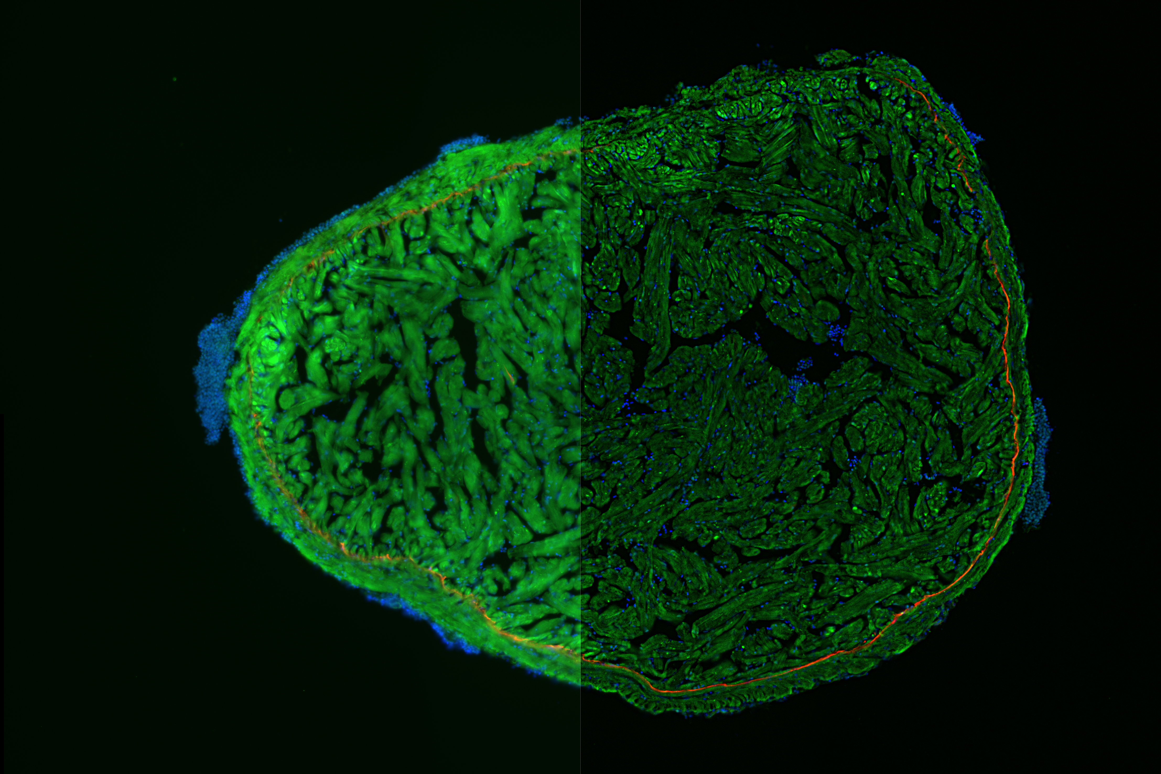
Kidney Section - THUNDER Imager 3D Tissue
Retina Section - THUNDER Imager 3D Tissue
Heart Section - THUNDER Imager 3D Tissue
Liver Section - THUNDER Imager 3D Tissue
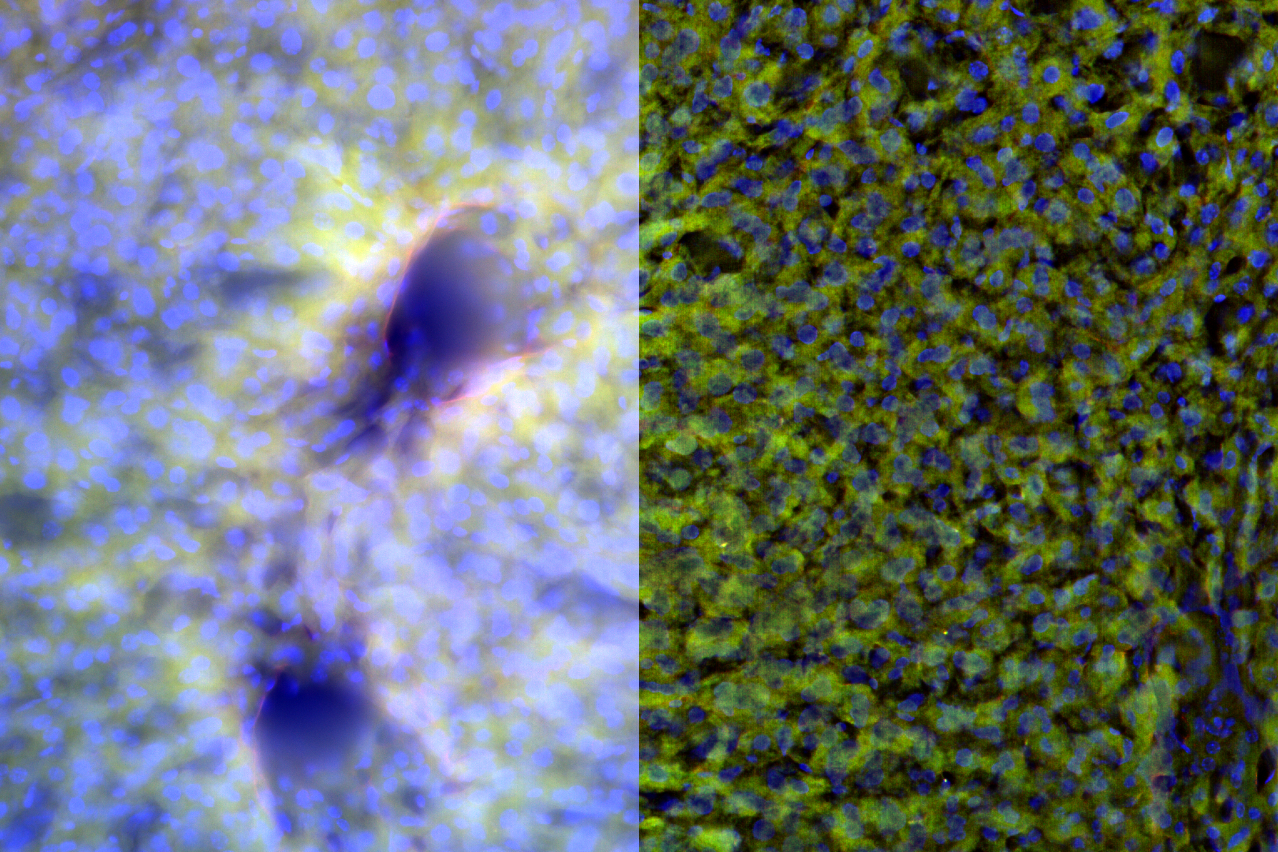
Image courtesy of Dr. Remy Bonnavion, Max Planck Institute for Heart and Lung Research, Bad Nauheim, Germany
Brain Section - THUNDER Imager 3D Tissue
Mouse Retina - THUNDER Imager 3D Cell Culture
Sample courtesy by Jeremy Burton, PhD and Jiyeon Lee, PhD, Genentech Inc., South San Francisco, USA. Imaged by Olga Davydenko, PhD (Leica)
Mouse Aorta - THUNDER Imager 3D Tissue
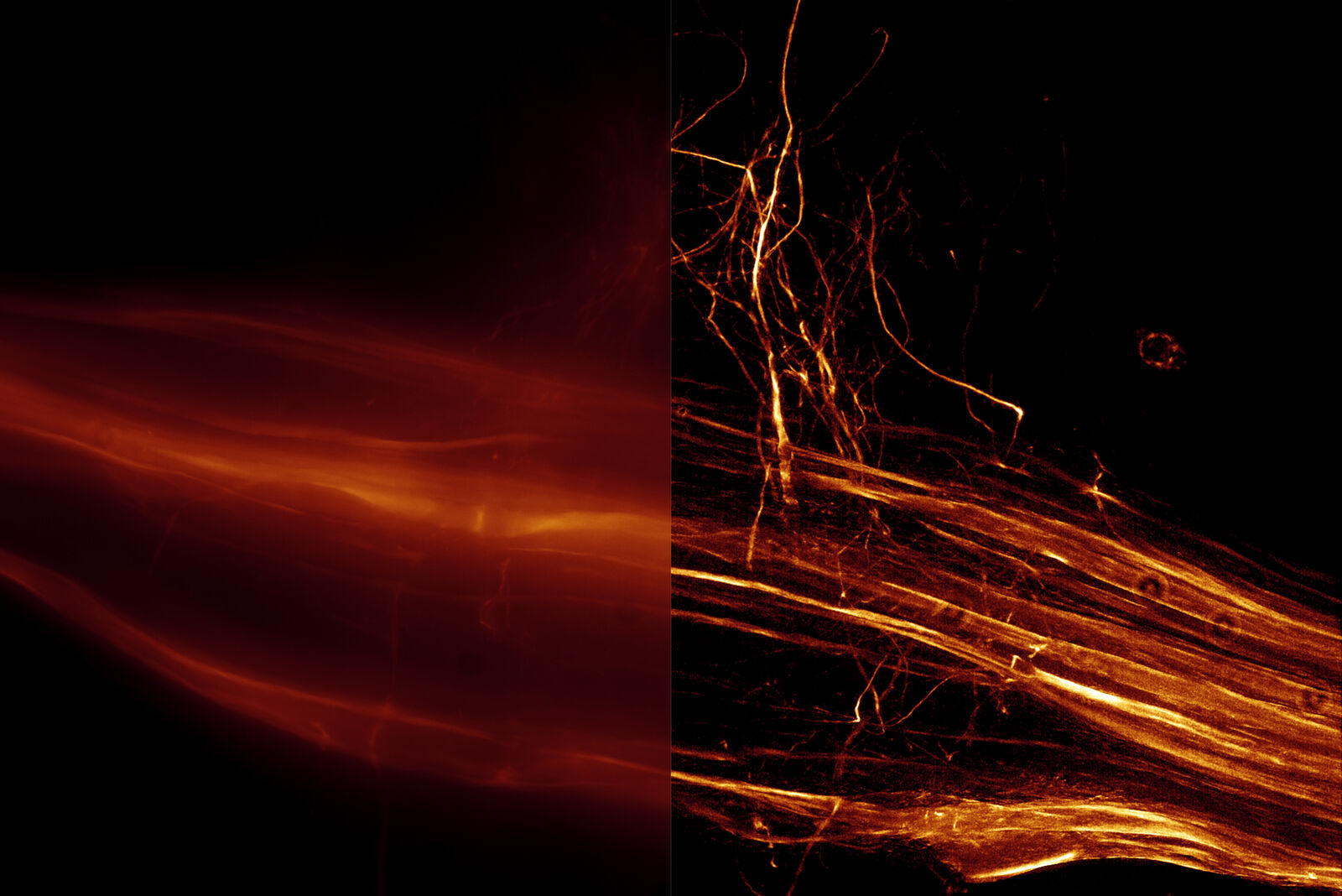
Fiber - THUNDER Imager 3D Cell Culture
Mouse Lens Section - THUNDER Imager 3D Cell Culture
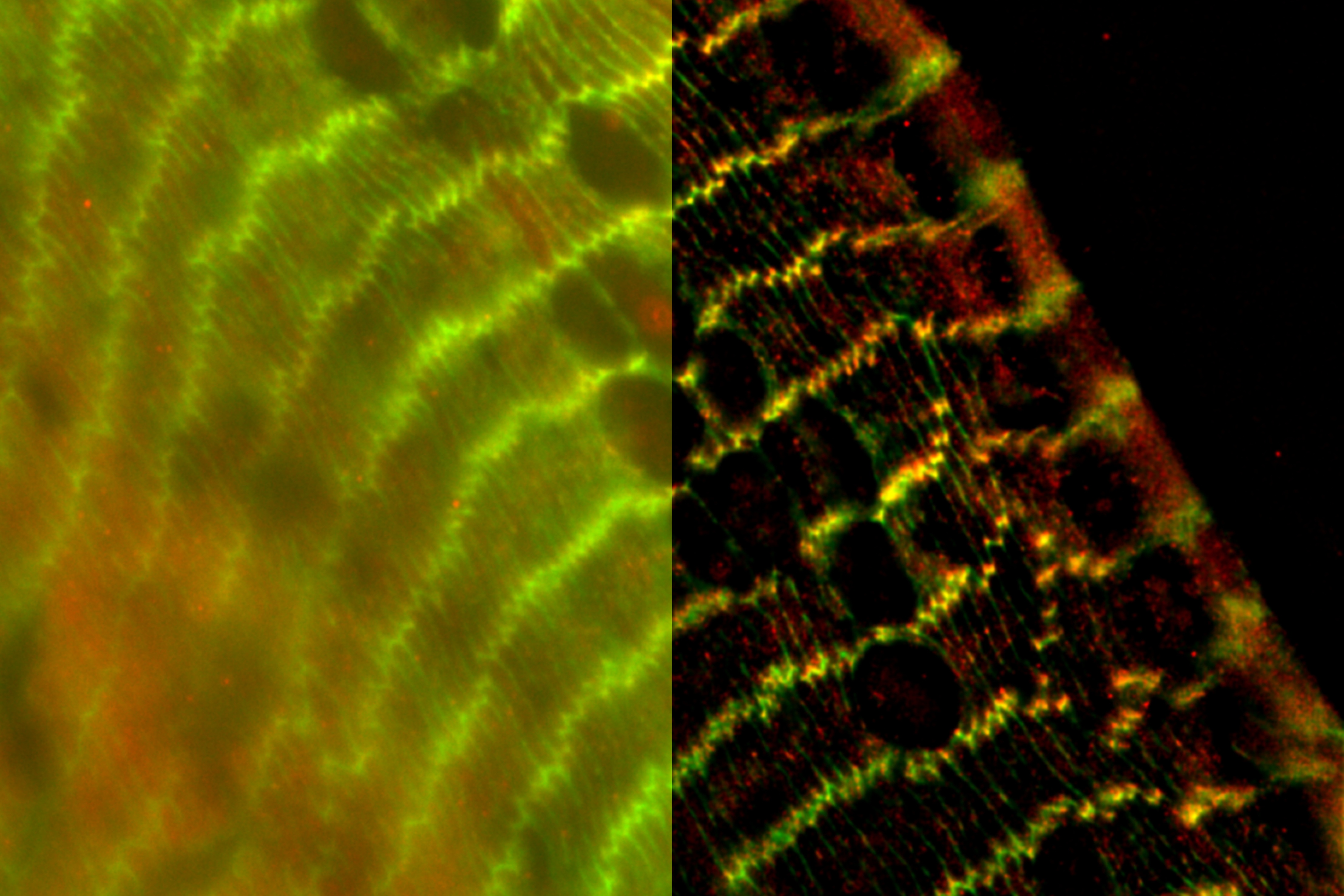
Image courtesy of Dr. Nathalie Houssin, Plagemen Lab, Ohio State University, USA
Human hepatic progenitor cell (HPC) - THUNDER Imager 3D Tissue
Mouse lung - THUNDER Imager 3D Cell Culture
Drosophila Follicle - THUNDER Imager 3D Cell Culture
Brine Shrimp leg - THUNDER Imager 3D Cell Culture

Image acquisition performed with DMi8, HC PL APO 63x/1.40 Oil Objective and DFC9000 GT camera.
Image size 2048 x 2048 pixels, 274 z-sections.
Image taken by Louise Bertrand, Leica Microsystems, USA.
Related Articles
-
, tubulin with Cy5 (red), and nuclei with DAPI (blue). Image courtesy of Dr. Günter Giese, Max Planck Institute for Medical Research, Heidelberg, Germany.")
Overview of Fluorescent Dyes in terms of Applications and Properties
An introduction to commonly used fluorescent dyes and an overview of their characteristics are given…
Mar 16, 2026Read article -
Factors to Consider When Selecting a Research Microscope
An optical microscope is often one of the central devices in a life-science research lab. It can be…
Dec 16, 2025Read article -

Technical Terms for Digital Microscope Cameras and Image Analysis
Learn more about the basic principles behind digital microscope camera technologies, how digital…
Dec 14, 2023Read article
