Related Articles
-

Improving Zebrafish-Embryo Screening with Fast, High-Contrast Imaging
Discover from this article how screening of transgenic zebrafish embryos is boosted with high-speed,…
Jun 16, 2025Read article -

Unlocking the Secrets of Organoid Models in Biomedical Research
Get ready to delve deeper into the world of organoids and 3D models, which are essential tools for…
May 19, 2025Read article -
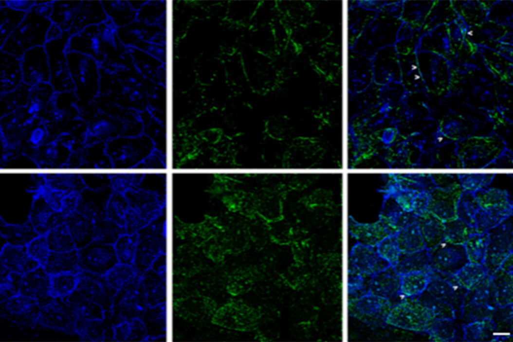
 applied. Image courtesy of Samuel East, Uncommon Bio.")
Designing the Future with Novel and Scalable Stem Cell Culture
Visionary biotech start-up Uncommon Bio is tackling one of the world’s biggest health challenges:…
Mar 11, 2025Read article
