STELLARIS 공초점 현미경 플랫폼
차세대 STELLARIS는 연구원들의 필요에 맞춰 개발한 공초점 현미경 플랫폼입니다.
STELLARIS 공초점 현미경은 FLIM, STED, DLS 및 CRS를 포함한 모든 라이카 장비와 결합할 수 있습니다. 차세대 STELLARIS 공초점 플랫폼으로 더 쉽고 빠르게 보람 있는 연구 결과를 얻어보세요. 강력하며 가능성과 생산성을 높입니다
더 많이 발견할 수 있는 가능성
STELLARIS의 특별한 TauSense 기술을 통해 모든 시료에서 추가적인 막 정보를 추출하고 연구의 과학적 영향력을 높일 수 있습니다. TauSense는 형광 수명을 이용하는 응용 분야를 위한 영상촬영 도구로 구성되어 있어 세포 맥락 안에서 분자 기능을 탐색할 수 있습니다.
- TauContrast는 신진대사 상태, pH, 이온 농도 등의 기능적 정보를 즉시 제공합니다.
- TauGating은 원치 않는 형광 잡음을 제거하여 이미지 품질을 개선합니다.
- TauSeparation은 스펙트럼 옵션을 넘어 실험에서 형광 신호의 조합을 확장합니다.
- TauInteraction은 분자 상호작용(예: 단백질-단백질 상호작용)의 간단한 검출 및 정량화를 제공합니다.
더 많은 작업이 가능한 생산성
간소화된 설정 및 촬영: STELLARIS 스마트 사용자 인터페이스인 ImageCompass는 사용자가 몇 번의 클릭만으로 고도로 복잡한 실험을 쉽고 직관적으로 설정할 수 있는 방법을 제공합니다.
- 빠르고 간편함: Aivia가 제공하는 LIGHTNING 초고해상도 동적 신호 향상(Dynamic Signal Enhancement, DSE)을 통해 실시간으로 최대 속도로 뛰어난 이미지 품질을 제공합니다.
- 직관적인 사용자 인터페이스: ImageCompass는 실험 설정부터 촬영까지 사용자를 지원합니다.
- 실험 최적화: LAS X Navigator와 같은 도구가 매끄럽게 통합되어 간단한 이미징이 가능합니다.
중요 데이터에 액세스하기
Aivia의 자율 현미경으로 고품질 결과를 더 빠르게 얻으세요.
- STELLARIS의 생명 과학용 AI 기반 희귀 이벤트 감지 워크플로우는 생물학적 샘플에서 희귀 이벤트의 최대 90%를 자율적으로 감지합니다.
- 관심 있는 물체만 식별 및 기록되므로 데이터 획득 시간을 최대 70%까지 단축합니다. 필요한 것만 기록하여 하드 드라이브의 귀중한 공간을 절약합니다.
- 현미경 검사에 소요되는 시간이 줄어듭니다. 자율 희귀 이벤트 감지 워크플로우는 일반적으로 소요되는 시간을 대폭 줄입니다.
- Aivia가 지원하는 자율 현미경으로 시간 제약과 복잡성으로 인해 이전에는 불가능했던 실험을 수행해 보세요.
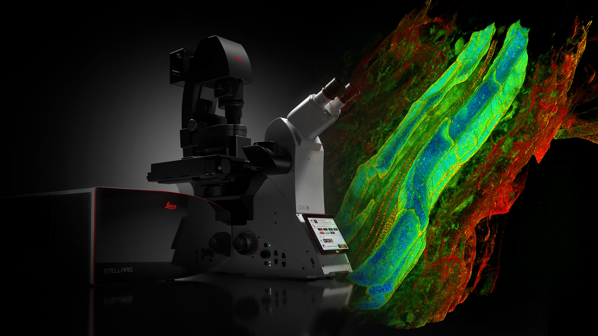
백색광 레이저가 장착된 STELLARIS
STELLARIS는 라이카의 독점 Acousto-Optical Beam Splitter(AOBS) 및 Power HyD 검출기에 백색광 레이저를 장착한 유일한 공초점 시스템입니다. STELLARIS는 특별한 TauSense 기술로 이미지 품질과 생성된 정보량에 대한 새로운 표준을 정립합니다.
백색광 레이저가 장착된 STELLARIS는 FAst Lifetime CONtrast(FALCON), STED, Deep In Vivo Explorer(DIVE), Digital Light Sheet(DLS), Cryo 및 CARS와 결합할 수 있습니다. STELLARIS는 이 장비들의 가능성을 극대화하고, 새로운 연구 표준을 정립할 수 있는 힘과 가능성을 선사합니다.
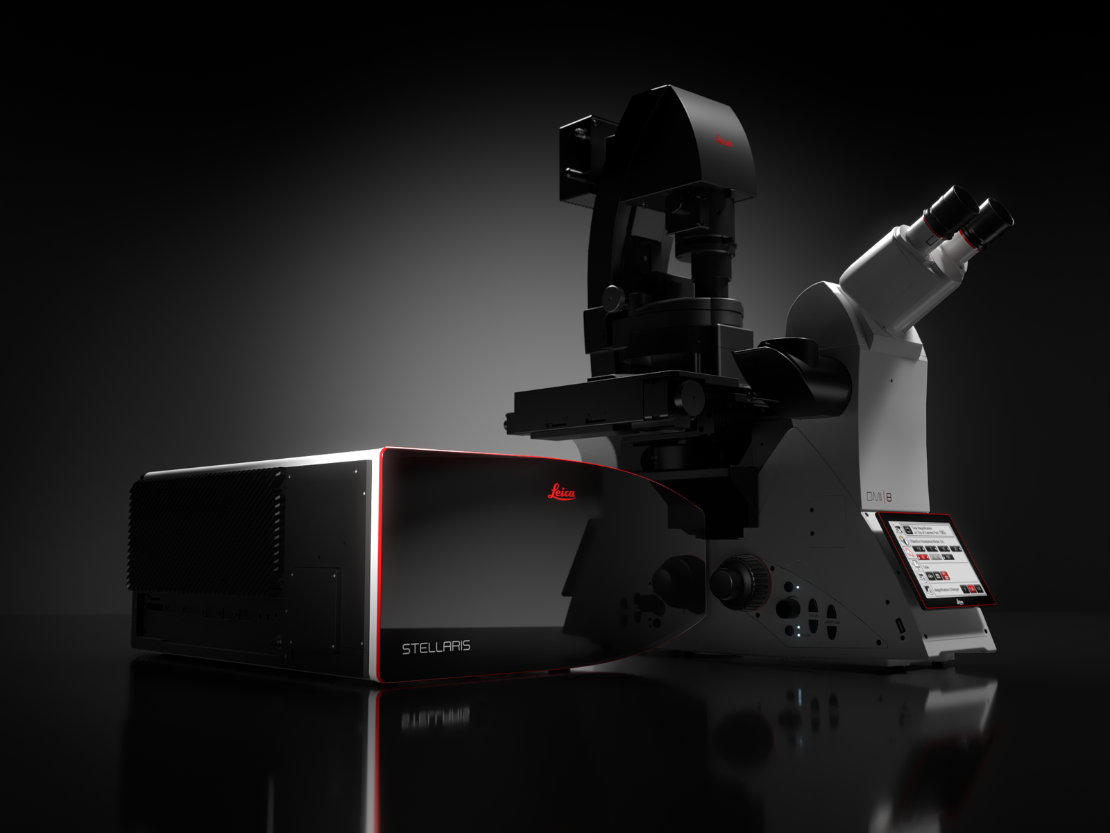
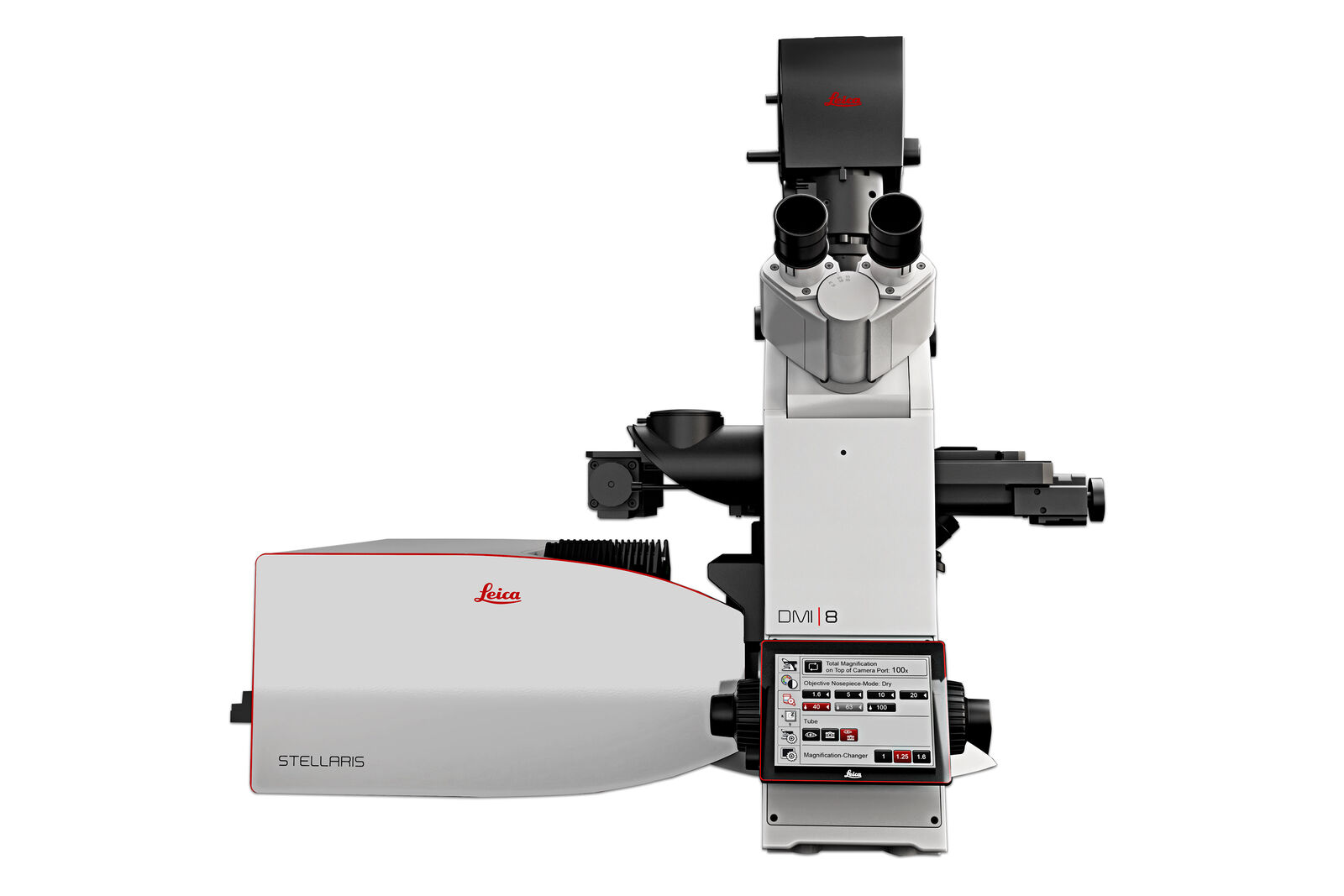
STELLARIS
STELLARIS는 스펙트럼 검출, 광자 계수 검출기 및 공초점 초고해상도를 제공합니다.
실험 설정 및 촬영 과정을 직관적인 방식으로 안내하는 스마트 사용자 인터페이스 ImageCompass를 사용해 쉽게 다채널 공초점 영상촬영을 수행할 수 있습니다.
더 많은 것을 보는 힘
Power HyD 검출기 제품군은 더 높은 광자 검출 효율성(PDE)*, 매우 낮은 다크 노이즈 및 410~850nm의 민감한 스펙트럼 검출 기능을 제공합니다.
- 뛰어난 이미지 품질: 밝기, 해상도 및 대비의 이상적인 조합.
- 완벽한 스펙트럼 자유도: 라이카의 차세대 백색광 레이저를 사용하면 스펙트럼 전체에서 최대 8개의 단일 여기 라인을 동시에 사용할 수 있습니다. 다른 공초점 플랫폼보다 더 많은 형광단 조합을 이미징하고 더 많은 라벨을 병렬로 사용하여 옵션을 NIR 범위로 확장할 수 있습니다.
- 살아있는 세포의 섬세한 이미징: 필요한 최저 조명 수준에서 효율적인 신호 획득을 통해 샘플의 무결성과 이미지를 장기간 유지합니다.
*기존의 다중알칼리 광전증폭관 튜브(PMT)에 비해 광자 검출 효율성(PDE)이 2배 더 높고, 확장된 빨간색 범위에서는 3배 더 높습니다.