
TauSense: a set of imaging tools that give users instant access to fluorescence lifetime information at the push of a button
Leica Microsystems’ TauSense technology is a set of imaging tools based on fluorescence lifetime. Found at the core of the STELLARIS confocal platform, it will revolutionize your imaging experiments. Whatever your sample or staining procedure, fluorescence lifetime information is always there. Now TauSense gives you access to this additional information and expands the potential of your research with the possibilities provided by different TauSense modes.
Adding an extra dimension of information to your experiments?
To get the most from your precious samples and expensive reagents, it is important to maximize the amount of information you obtain from each and every experiment. Advances in microscopy technology allow us to see our samples in different ways. New methods can be explored to give new, clear insights and to see things that have never been seen before.
Best of all, the lifetime parameter is an intrinsic characteristic of fluorescent molecules, so lifetime data is available “for free” with every fluorescence experiment.
Study the effect of microenvironment on events using TauContrast
Changes in the cellular microenvironment can affect the fluorescence lifetime of fluorophores. When this happens in a predictable way, it is possible to use fluorophores as biosensors.
Many fluorophores have lifetimes that vary depending on environmental conditions such as pH or ion concentration. The TauSense mode TauContrast provides immediate access to additional functional information such as metabolic status, pH and ion concentration. TauContrast generates useful experimental images with lifetime-based contrast where each pixel contains information about the average photon arrival time as well as the fluorescence intensity (photon count). This makes it easy to visualize changes in the microenvironment as pixels can be differentiated with different arrival times and therefore produce an extra contrast map based on the effect of the microenvironment (e.g. high calcium vs. low calcium conditions).
For example, the concentration of Ca2+ ions affect the lifetime of Oregon Green [1]. Using TauContrast, the photon average arrival times of Oregon Green can therefore give a readout of the calcium concentration. As shown in Figure 1, this is particularly useful for studying calcium waves in a variety of cell types or for visualizing dynamic changes in calcium concentration and calcium waves in live cells.

Another example is fluorescein, which exhibits changes in lifetime with respect to pH changes in the local microenvironment. Fluorescein can be added to nanoparticles as a pH sensor and to monitor the permeability of vesicular membranes. Nile red is yet another useful dye for biosensing. By observing lifetime changes of Nile red, it is possible to measure intracellular lipid polarity, which varies between distribution of lipid droplets and lipid rich regions of the cell [2].
Multiplex beyond fluorescence spectra
Technical limitations in the past have made selecting dyes to optimize confocal imaging overly complex and time-consuming. This is especially true in experimental setups where staining procedures, overlapping fluorescent emission from different dyes and autofluorescence can potentially mask important signals, and its removal require a lot of careful and time-consuming optimization work. TauSense is now putting more experimental flexibility and potential into the hands of researchers, so that they can extract more information from every sample.
Separation of spectrally overlapping dyes with TauScan and TauSeparation
Crosstalk between spectrally similar fluorophores can completely mask valuable information in your experiment. However even if the spectra of two fluorophores overlap completely, it is possible that signals from each one can be distinguished exploiting their differences in fluorescence lifetime. TauScan and TauSeparation make use of fluorescence lifetime-based information to separate fluorophores that could not previously be separated with spectral unmixing by exploiting the lifetime differences to distinguish if the photons are emitted from one probe or another.
The TauSense mode TauScan works by calculating the mean lifetime components of your samples’ fluorescence and displays them as a distribution curve, enabling you to determine the mean lifetime components of the different probes in the sample. This helps you divide the entire range of detectable lifetime components and assign them to different species in your sample.
When different fluorescent mean lifetime components have been determined with TauScan, the TauSense mode TauSeparation can be used to separate species with overlapping spectra based on their differing lifetimes. TauSeparation splits the probes present in the sample based on their fluorescent mean arrival component and records the resulting images in separate channels.
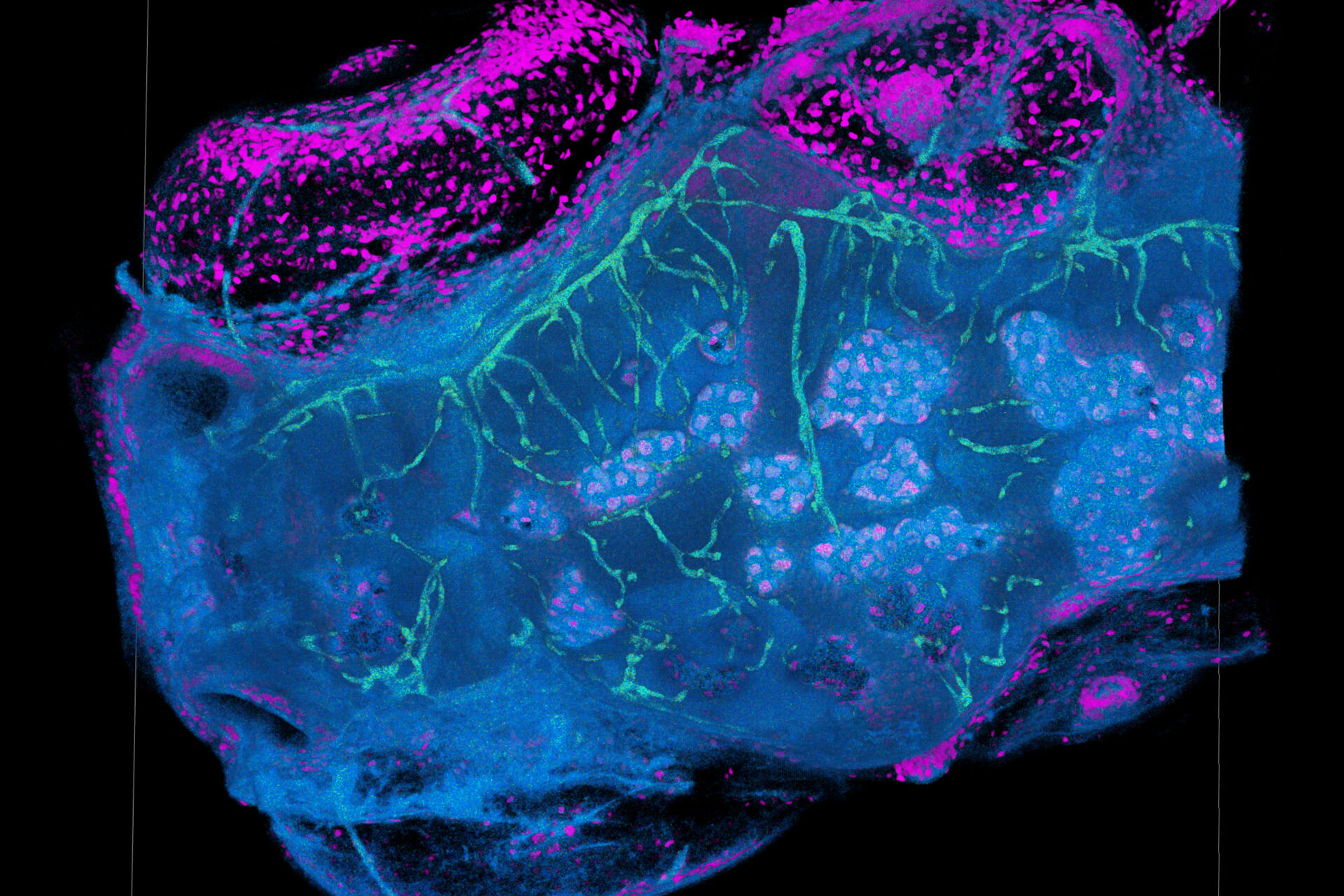
The potential to separate dyes species using their lifetime signatures is particularly important in live cell and in vivo imaging, where limitations in the number of available fluorescent probes that can be distinguished in a single experiment reduce the amount of information you can get from a single experiment [3]. As displayed in Figure 2, dyes with very similar spectra such as mNeonGreen and Mitotracker green or SiR and NucRed can now be easily distinguished thanks to their different fluorescence lifetime signatures.

TauSeparation even when their intensities overlap.
Improving confocal image quality
Increasing the quality of image data is the goal of every confocal microscopy user. High quality means removing background signals and maximizing detection efficiency preserving the desired signal in your sample. This is a strong advantage of TauSense.
Removing unwanted fluorescence contributions using TauSense Gating
Many tissues exhibit endogenous autofluorescence from naturally fluorescing molecules such as aromatic amino acids or cytokeratins.
With STELLARIS, eliminating unwanted fluorescence background, such as autofluorescence or reflections is made possible with the TauSense gating modes (TauGating and GateScan) which enable you to set digital gates for the easy elimination of unwanted signals from your images. Gating can separate fluorescence signals with short lifetimes, as it is the case with some autofluorescence signals. TauGating works by separately recording channels in different time windows where photons are detected. While all windows are combined to form an intensity image, you can eliminate those that are a contribution from your unwanted signal, lifting a veil from the image to reveal the true signal beneath.
As an example, autofluorescence in plants can interfere with the signal from fluorophores and limit the amount of information that can be obtained from an experiment. It is possible to quickly scan and remove background autofluorescence with short lifetimes such as the signal from autofluorescencing organelles like chloroplasts.
 from the desired fluorescent signal from the cell membrane (green).")
Summary and conclusion
TauSense is a toolbox for confocal microscopy users aimed at putting the potential of lifetime information into a familiar setup. TauSense gives you access to an extra dimension of information, expanding the potential of your confocal experiments and maximizing the information you get from your sample. In short, TauSense brings the potential of fluorescence lifetime to biologists.

, actin network (ATTO 647N), and nuclear pore basket (CF 680R).")
