Surgical Microscopes for Spine Surgery
Leica Microsystems surgical microscopes enhance precision and efficiency, especially in minimally invasive spine surgery, by delivering advanced visualization, ergonomic comfort, and seamless OR integration, ensuring uninterrupted, high-resolution views tailored to the unique demands of modern spine procedures.
Contact a local specialist for expert advice on the right solution for your needs and budget.

Why is clear visualization crucial during spine surgery?
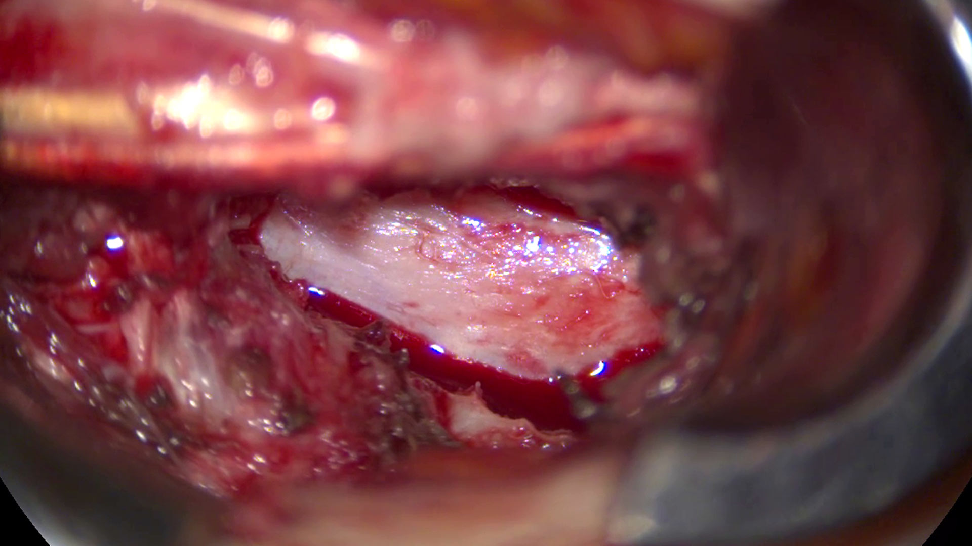
Clear, bright visualization is critical in spine surgery, where surgeons work near the spinal cord, nerve roots, and dura during lumbar and cervical decompression, discectomy, pedicle screw placement, spinal fusion, and minimally invasive spine surgery (MISS).
Compared to loupes, optical magnification and high-quality illumination enable precise tissue differentiation, decompression, and safer neural handling.

Which tool improves visualization during spine surgery?
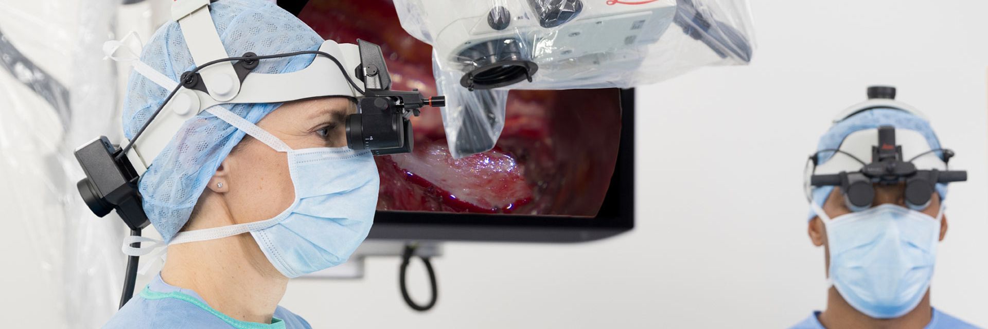
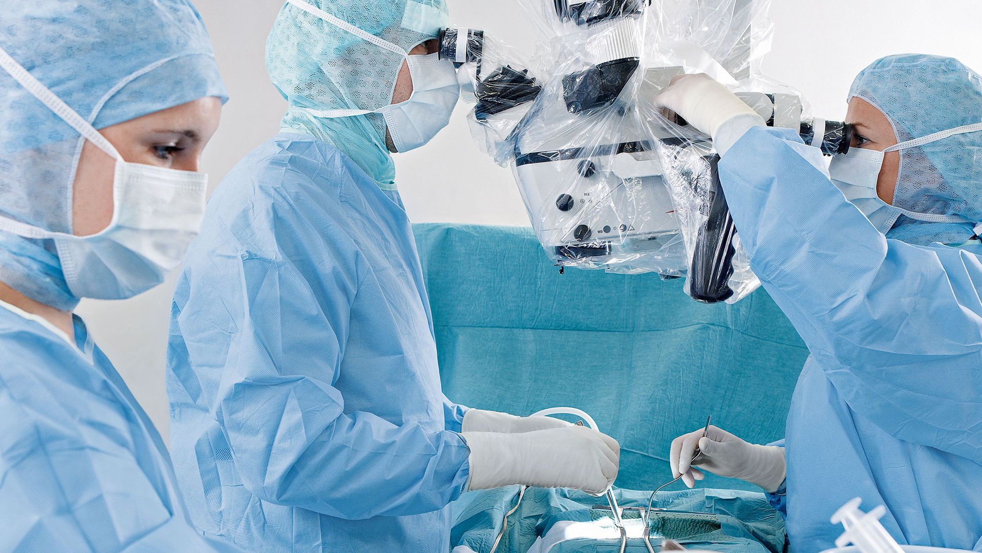
Surgical microscopes deliver advanced visualization in spine surgery, offering high magnification, resolution, depth of field, and superior illumination for precise work in deep or complex approaches. Compared with surgical loupes, microscopes provide better image quality and depth perception, improving visualization of small anatomical structures.

How can advanced illumination support spine surgery?
Advanced illumination supports spine surgery by providing bright, coaxial illumination that minimizes shadows in deep and narrow cavities. Microscope-based illumination ensures consistent brightness even at high magnification, and minimizes shadows caused by instruments or anatomical depth. This enhanced illumination improves the identification of critical structures such as nerve roots, the dural sac, and disc fragments.

Does ergonomic strain differ when using microscopes?
Loupe-based spine surgery often requires sustained neck flexion, increasing cervical spine loading and musculoskeletal strain. In contrast, surgical microscopes with 3D heads-up viewing technology support an upright, neutral posture, reducing physical stress, discomfort, and fatigue while improving endurance and ergonomics during spine surgery procedures.
What makes Leica surgical microscopes an excellent choice for spine surgery?
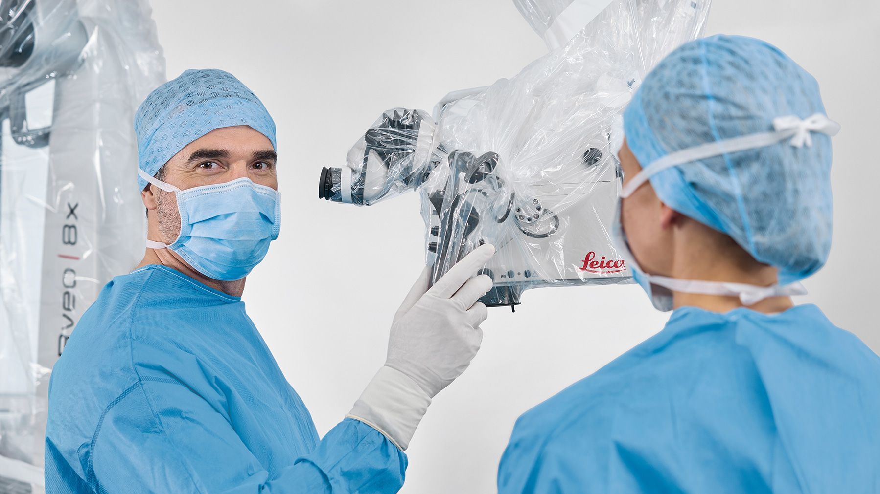
Maneuverability & Positioning Freedom
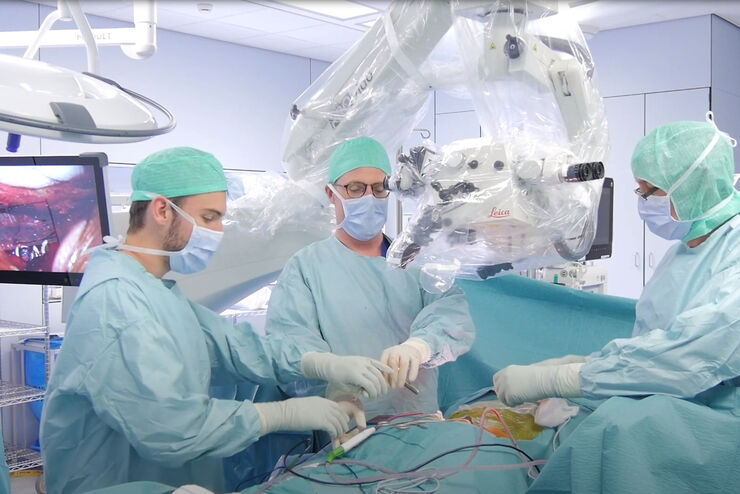
With internally routed cables, large overhead clearance, extensive long arm reach, electromagnetic brakes and a compact optics carrier design, maneuvering is smooth and effortless with Leica surgical microscopes. Their 600 mm working distance allows for easy handling and passing of instruments.
High quality optics & Illumination
Premium optics deliver crisp images with enhanced depth perception for accuracy and precision during microsurgery. Powerful xenon illumination provides a clear view with natural colors deep into the operating field. Small Angle Illumination reduces shadows in narrow cavities, and built-in controls help during surgery.
Ergonomics & Efficiency
Our microscopes support ergonomics in spine surgery with 360° rotatable binoculars, full range of movement and tilt of the optics carrier and fast focusing to keep interruptions to a minimum. Optional 3D visualization further boosts surgical ergonomics, especially during long procedures with challenging patient positioning.
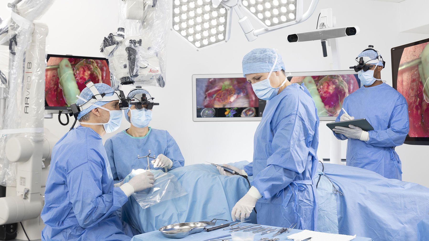
Training & OR Collaboration
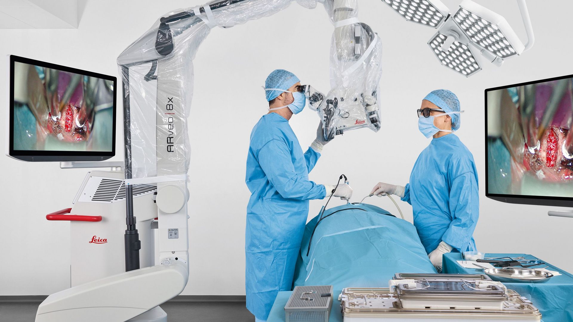
With the ARveo 8x 3D digital microscope, surgeons and the entire OR team can conduct and observe surgery with 3D depth perception onscreen or via the MyVeo headset. 3D visualization eases anatomical and spatial understanding and allows everyone to follow the surgical course easily and in real time.
Our Solutions for Spine Surgery
ARveo 8x | M530 OHX | PROvido | |
| MyVeo all-in-one surgical visualization headset | Compatible | Non-compatible | Non-compatible |
| Heads-up surgery | 55" 3D 4K monitor for heads-up surgery | 2D view only for observation | 2D view only for observation |
| 3D 4K Visualization | Available | Not available | Not available |
| RemoteCare: Smart monitoring with a cloud-based service | Available | Not available | Not available |
| FusionOptics technology | Available | Available | Available |
| +40% Magnification multiplier | Available | Available | Available |
| Video recording | 3D 4K, 2D 4K | HD | HD |
| Working distance | 600 mm | 600 mm | 600 mm |
| Go to product page | Go to product page | Go to product page |
Related Articles
 case.")
Flexibility and Efficiency in Minimally Invasive Spine Surgery
Advanced Visualization: Transforming Minimally Invasive Spine Surgery

Use of AR Fluorescence in Neurovascular Surgery
Minimally Invasive Spine Surgery: Improving Precision and Accuracy with Microscopes
Frequently Asked Questions Spine Surgery
Surgical loupes are not optimal for deep or complex spine surgery. Loupes provide limited magnification and rely on external, non‑coaxial illumination, reducing visibility in narrow surgical corridors. Comparative studies show that, although outcomes may be similar in selected cases, microscopes offer superior visualization quality in minimally invasive lumbar fusion and decompression procedures.
According to the Manual of Spine Surgery, surgical microscopes are particularly beneficial in spine procedures that require precise visualization of neural structures within deep or narrow surgical corridors. These include lumbar and cervical discectomy and fusion, laminectomy for spinal stenosis, cervical myelopathy decompression, and spinal cord or vertebral tumor resection. In these surgeries, microscope-assisted visualization supports accurate decompression, controlled tissue removal, and improved safety around critical neural anatomy.
A large working distance is key in spine surgery for maneuvering long instruments.
The cost of a spine surgery microscope depends on the selected range: ARveo 8x with MyVeo, M530 OHX, or PROvido, and the required configuration. Factors such as digital applications, viewing options and video recording capabilities influence overall pricing across the portfolio.
