
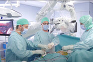
Role of GLOW800 during neurovascular surgery
With GLOW800 Augmented Reality fluorescence, Dr. Renner recounts, it is possible to see the angiographic picture in the microscope directly during neurovascular surgery, with an optimal depth perception. There is no need to recall and try to reconcile the black and white blood flow video with the natural anatomical view.
As such, GLOW800 provides real-time information about blood circulation and perfusion during the arterial and venous phase. This information allows to select the safest way and strategy in the treatment of a lesion. It also helps verify the effective and definite elimination of the lesion.
Neurovascular surgery: Brain & spine clinical cases
Dr. Renner presented 5 clinical cases:
- 2 aneurysm clinical cases: GLOW800 supports aneurysm clipping procedures and helps confirm that the aneurysm is excluded from the circulation. For optimal depth perception, it is important to have the right working distance and magnification.

- A spinal dural arterio-venous fistula clinical case: the patient was operated using GLOW800. The fistula was clipped and GLOW800 allowed to confirm that the fistula was definitely excluded from the circulation.

- A complex cranial dural arterio-venous fistula patient case: the treatment was combined, with surgical exposure using GLOW800 and one of the fistula points eliminated through intraoperative coiling.

- A low stroma hemangioblastoma patient case: the diagnosis was initially not clear and the surgeon removed a solid tumor to support the diagnosis. The procedure was performed with GLOW800 to visualize arterial inflow and identify the feeders.

GLOW800 provides real-time blood circulation and perfusion information within a controlled and tight workflow, without the need to turn away from the microscope.
Want to learn more? Register below to watch the full webinar presented by Dr. Christof Renner and see the clinical cases he shared.
Note: The statements of the healthcare professional in this video reflect only his opinion and personal experience. His statements do not necessarily reflect the opinion of any institution with whom he is affiliated. Please check with your local Leica Microsystems representative for product registration status in your region.
Related Articles
-
 case.")
Flexibility and Efficiency in Minimally Invasive Spine Surgery
According to Prof. Alex Alfieri, Chief Physician and Head of clinic for Neurosurgery and Spinal…
Jan 26, 2026Read article -

A Larger 3D Area in Focus for Neurosurgical and Ophthalmic Microscopes
Neurosurgeons and ophthalmologists deal with delicate structures, deep or narrow cavities and tiny…
Dec 18, 2025Read article -
Advanced Visualization: Transforming Minimally Invasive Spine Surgery
Over the past decade, spine surgery has evolved rapidly with the adoption of minimally invasive…
Dec 09, 2025Read article
