Structure and Physiology of Organoids and 3D Cell Culture
3D models range from simple multicellular cultures to complex models like organoids, which better mimic human tissues, enabling investigation of disease mechanisms and drug responses. Leica Microsystems provides precise, gentle, scalable solutions across 3D imaging workflows – from acquisition to analysis.
Our experts on solutions for Physiology of Organoids and 3D Cell Culture are happy to help you with their advice.

How can detailed structural analysis of organoids be achieved?
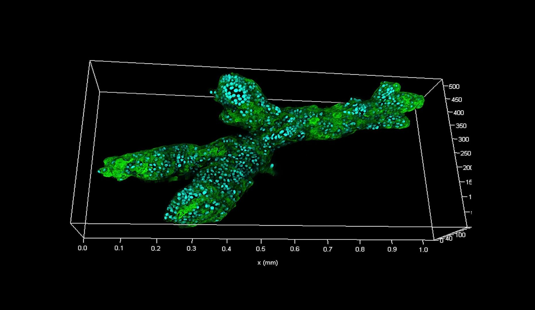
Using high‑resolution confocal imaging and light sheet microscopy, researchers can visualize and quantify intricate structures and details deep inside organoids and 3D cell cultures over time. Cleared sample imaging further helps visualize complex structures while 3D multiplexing enables parallel imaging of multiple cell types to better understand spatial relationships.

How can functional imaging reveal different aspects of organoid biology?
Functional imaging approaches such as FLIM and FRET based fluorescence lifetime imaging enable real time measurement of cellular processes in organoids, including protein interactions and signaling dynamics. These provide deeper insights into organoid biology, particularly when given spatial context by combining with structural imaging.

How can live cell dynamics be imaged in organoids and 3D cultures?
Imaging techniques such as advanced widefield, spinning disk confocal and light sheet microscopy enable real-time observation of live cells within 3D cultures. Fast, gentle imaging under physiologically-relevant conditions is essential to preserve cellular function, helping researchers study cellular dynamics and interactions and better understand native biological processes.
What are the advantages of Leica solutions for examining organoid structure and physiology?
Precise, high-resolution structural and functional imaging
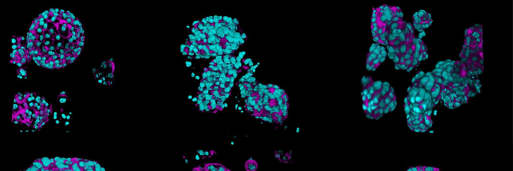
Complementary imaging approaches from Leica Microsystems enable detailed visualization of structures and processes deep within organoids. THUNDER Imager Cell Spinning Disk enables high speed, versatile imaging; the STELLARIS confocal microscope platform provides high resolution, multi modal flexibility; and Viventis Deep enables fast, gentle light sheet imaging of live and cleared samples over time.
Gentle live cell imaging to reduce photodamage
Light sheet microscopy enables gentle imaging of sensitive organoids and fast biological processes. By minimizing phototoxicity and photobleaching, Viventis Deep and Viventis SCAPE deliver gentle, long‑term, rapid volumetric imaging. With THUNDER Imager Cell Spinning Disk, diverse samples can be imaged at high-speed and high-resolution, with reduced phototoxicity.
Streamlined organoid imaging workflows
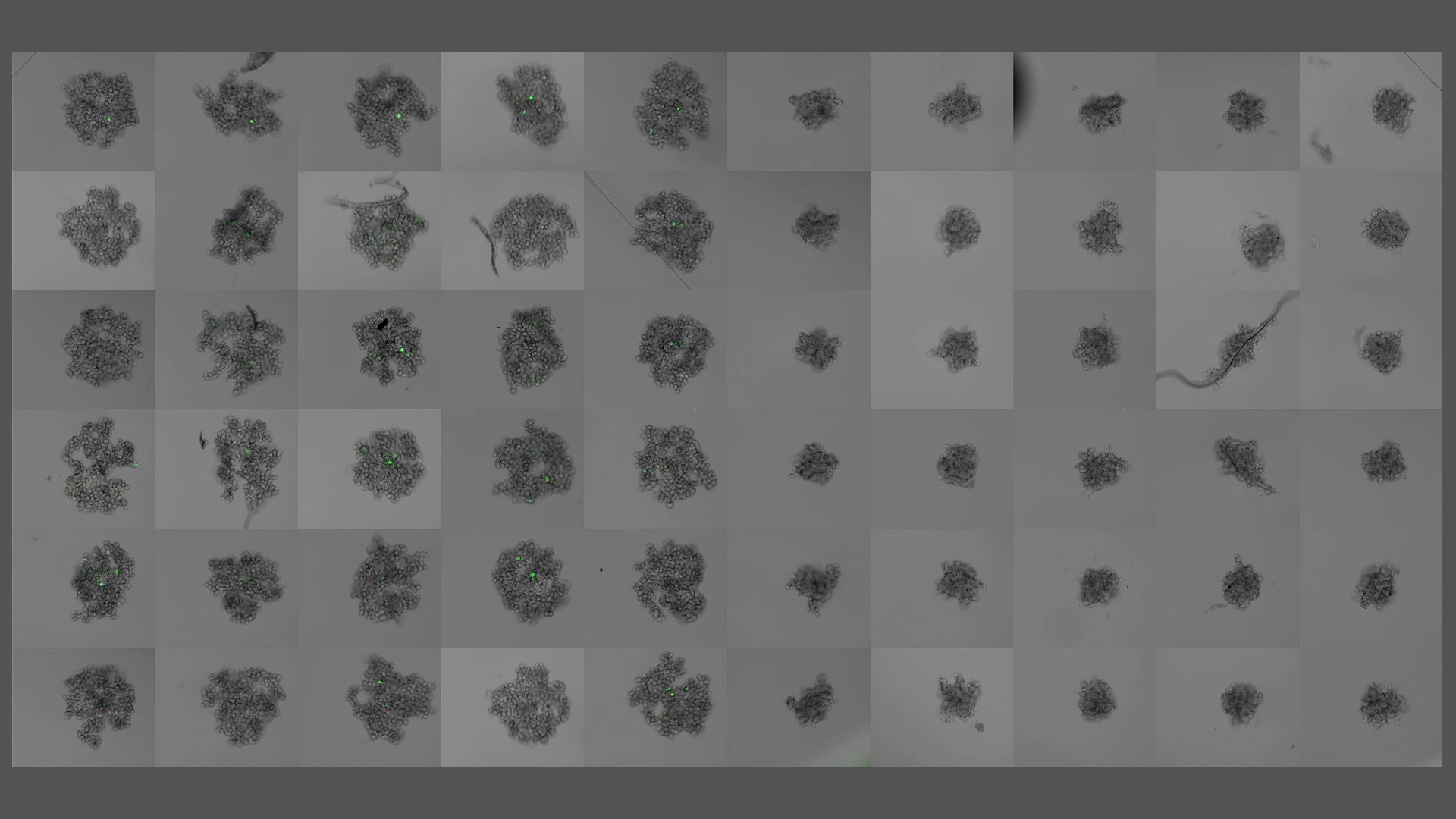
Monitor cell culture growth with a Mateo FL digital fluorescence microscope. Then pre‑screen organoids with THUNDER Imager Cell Spinning Disk before using Viventis light sheet microscopy for long‑term, gentle volumetric imaging. Complement with high‑resolution, high multiplex imaging using STELLARIS confocal microscopy. Then gain deeper insights with Aivia AI‑driven image analysis.
Advanced AI-powered analysis
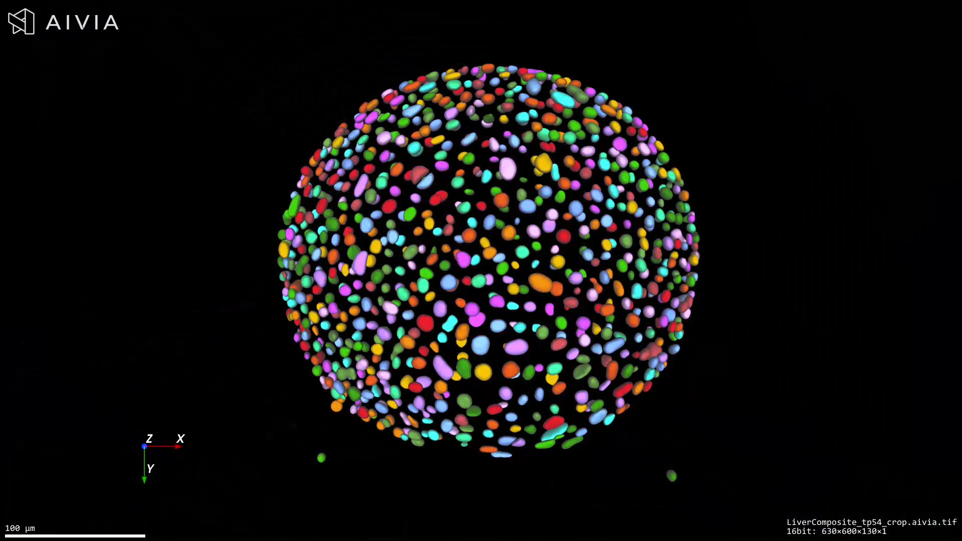
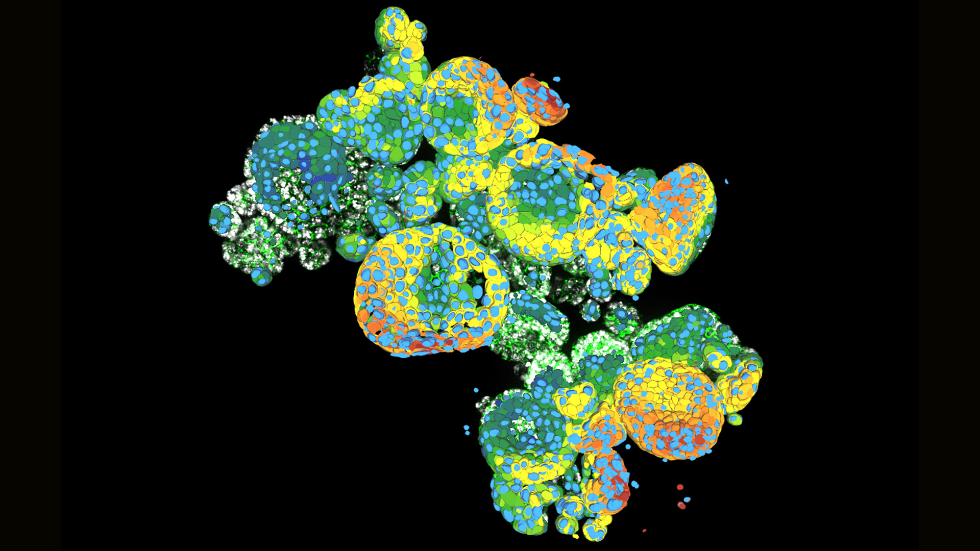
Aivia software offers a complete AI-based image analysis workflow from deep-learning cell segmentation to automatic phenotyping and data exploration in 3D samples. When using thick or highly scattering specimens, Aivia can be combined with THUNDER computational clearing technology, which removes out-of-focus-blur and gives cleaner data, resulting in better segmentation.
Frequently asked questions about Structure and Physiology of Organoids and 3D Cell Culture
3D cell cultures are systems where cells grow in three dimensions. Advanced models such as organoids can better mimic the structure and function of tissues in the body compared to traditional 2D cultures. 3D cell culture can range from simple multicellular structures such as kidney cell domes, through spheroids, to more complex structures such as organoids and assembloids.
Spheroids are simple clusters of broad-ranging cells that don't require a scaffolding to form 3D cultures and can't self-assemble or regenerate. Organoids are complex clusters of organ-specific cells, typically derived from primary tissue or grown in vitro from stem cells. They can self-assemble when given a scaffolding extracellular environment and represent the architecture and metabolism of their tissue of origin more closely than spheroids.
Assembloids are used to study interactions between organoids from different tissues, or to study co-cultures between organoids and different cell types such as T-cells or macrophages.
Imaging allows researchers to visualize cellular structures, monitor growth and differentiation, assess viability, and analyze responses to treatments in real time. It’s essential for validating experimental outcomes and gaining insights into complex biological processes.
There are several important considerations for 3D imaging, including:
- Penetration depth in thick 3D samples
- Speed of imaging to capture live events
- Image resolution
- Maintaining sample viability during live imaging (sensitivity)
- Long-term volumetric imaging
- Gentle sample handling of delicate 3D cultures
- Requirement for non-iterative multiplexing
- Analyzing large complex image datasets
Common techniques include:
- Light sheet microscopy
- Confocal laser scanning and spinning disk microscopy
- Widefield/epifluorescence microscopy
- Multiphoton microscopy
- Brightfield and phase contrast microscopy
- Label-free imaging
Each method offers different advantages in terms of resolution, depth, speed, etc. so it is important to consider the individual application before deciding which approach is best. At Leica Microsystems, our friendly expert team will be happy to assist you with this.
Imaging live cells provides insights into how cells behave in a more physiologically relevant way. In organoids and 3D cultures, live imaging helps track development, differentiation, and responses to stimuli or treatments without disrupting the sample.
Advanced image analysis software can quantify features such as cell morphology, proliferation, marker expression, and spatial organization. AI and machine learning tools are increasingly used to automate and enhance analysis.
NAMs (New Approach Methodologies) are non‑animal, human‑relevant research methods used in biomedical research. These methods include advanced technologies such as organoids, organ‑on‑a‑chip systems, and in‑vitro human cell models, often combined with computational or AI‑based analysis. They aim to improve biological relevance and reduce animal use.
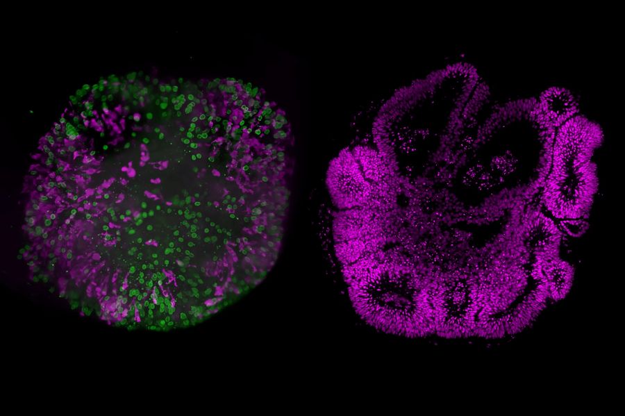
Related Articles
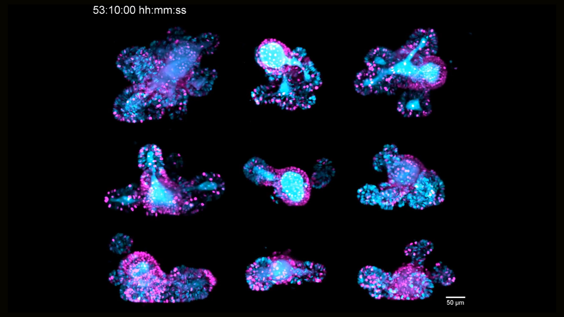
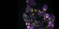
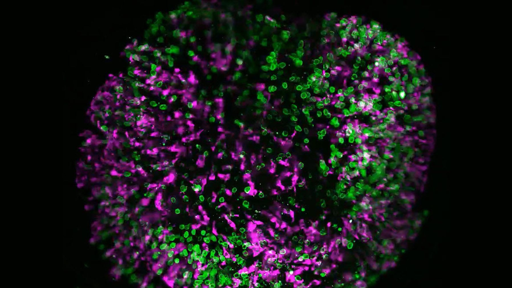
 and phalloidin (magenta), imaged using Viventis SCAPE; scale bar 50μm. Courtesy of Marina Cuenca and Heleen Jungen (Dayton lab), EMBL Barcelona.")
What’s the Best Organoid Imaging Approach for Early Drug Discovery?
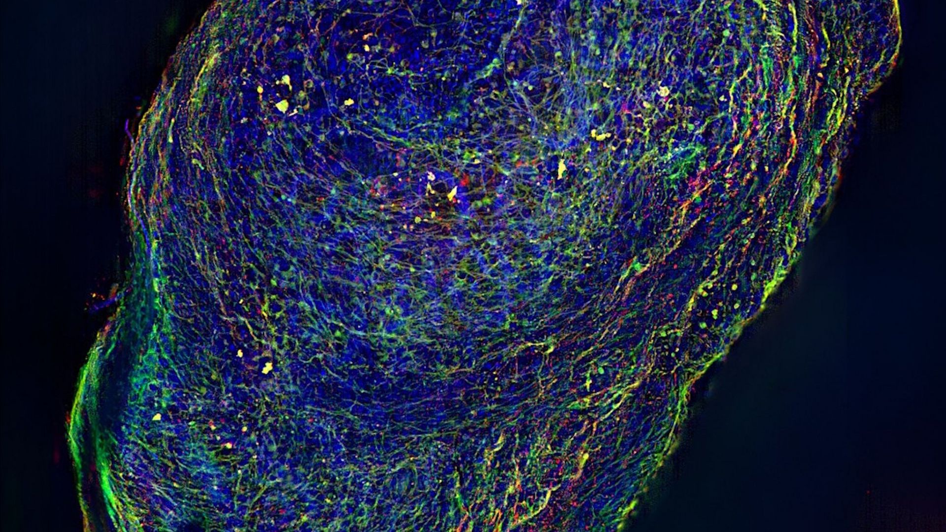
 2D slices of a 1 mm diameter midbrain neural organoid stained with DAPI (blue, nuclear stain), β-tubulin (green, neuronal stain), and GFAP (red, astrocyte stain).")
Fast, High-Contrast Widefield Imaging of Optically Challenging Samples

Multiscale Imaging of Organoids: High Content to Light Sheet

History, Developments and Trends of Microscopy in Cancer Research
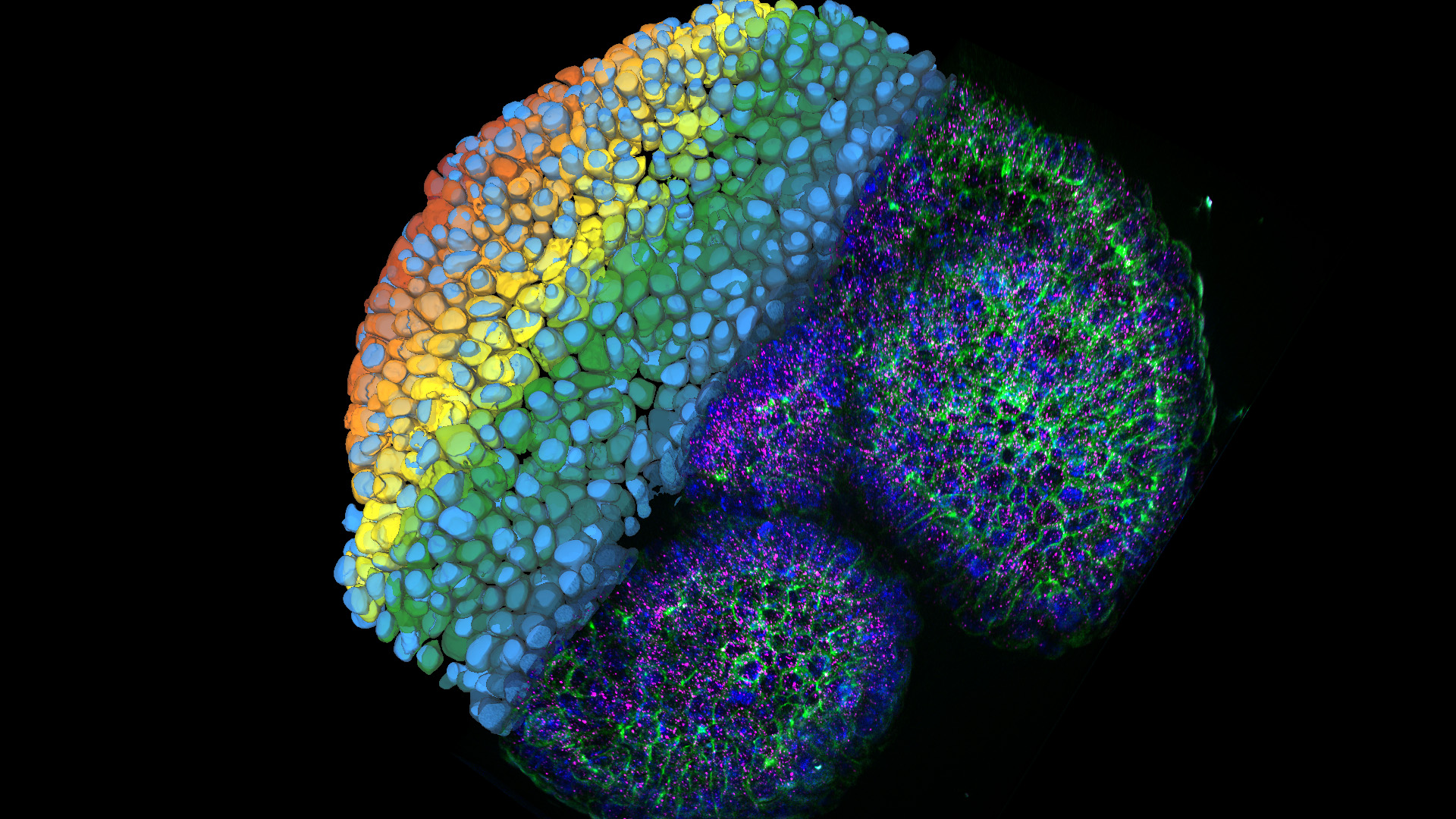
, tubulin with Cy5 (red), and nuclei with DAPI (blue). Image courtesy of Dr. Günter Giese, Max Planck Institute for Medical Research, Heidelberg, Germany.")
Overview of Fluorescent Dyes in terms of Applications and Properties

Researchers Insights: Microscopy in Cancer Research
.")
Focus on Long-Term Imaging in 3D with Light Sheet Microscopy
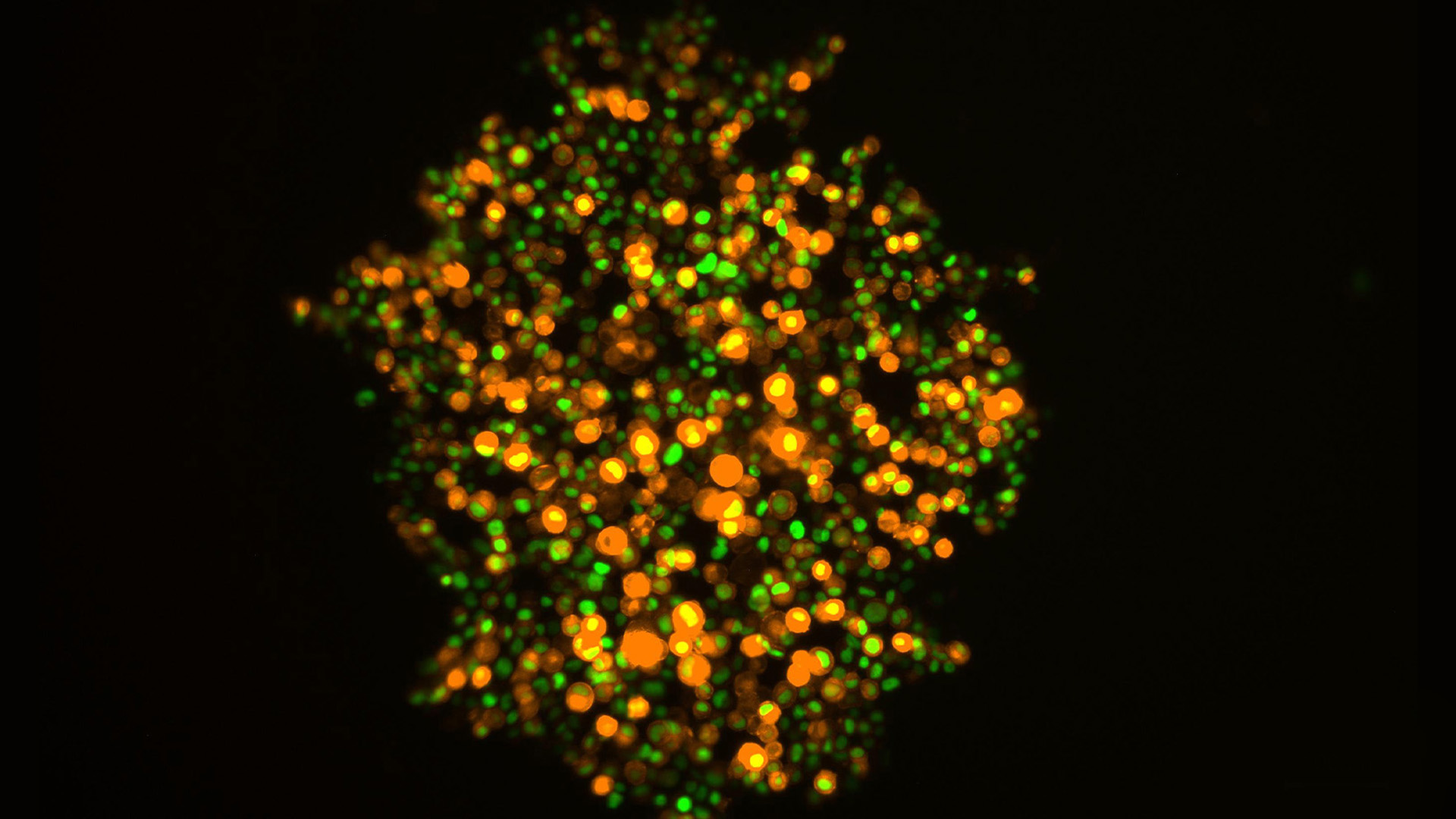
 at 2 weeks. Image acquired using Mica.")
How to Image Axon Regeneration in Deep Muscle Tissue

Capturing Developmental Dynamics in 3D

Integrated Serial Sectioning and Cryo-EM Workflows for 3D Biological Imaging
Organoids in New Approach Methodologies
The FDA’s Modernization Act 2.0 aims to reduce reliance on animal models by shifting toward human-relevant systems in preclinical workflows.
Organoids are increasingly used in new approach methodologies (NAMs) for biomedical research and drug discovery*.
Benefits of organoids include:
- Lower cost than animal models
- Higher scalability for faster results
- Less ethical constraints
- More accurate, predictive insights
- Fewer official licenses required
* Zushin et al. (2023). FDA Modernization Act 2.0: transitioning beyond animal models with human cells, organoids, and AI/ML-based approaches. J. Clin. Invest, 133 (21); e175824. https://doi.org/10.1172/JCI175824
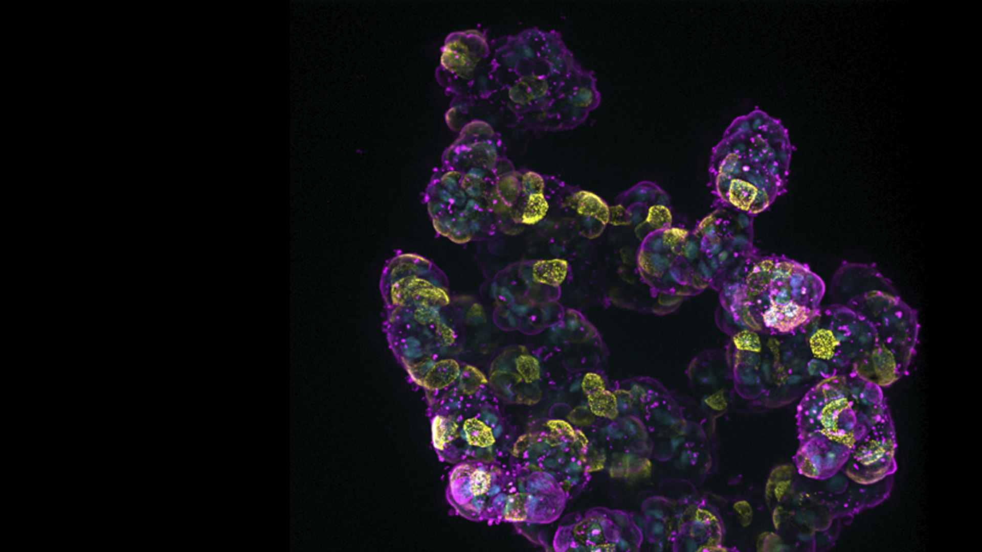
Organoids play an important role in our work, allowing us to model tumors in 3D and explore behaviors of engineered T cells in a way that’s both meaningful and patient specific. Imaging is central to this as we aim to understand how cancer and immune cells communicate at their most intricate level, where every cell, structure, and interaction within a tumor can be visualized, understood, and ultimately targeted.
We use advanced microscopy platforms from Leica Microsystems to explore cellular dynamics and spatial interactions. These include STELLARIS 8 FALCON FLIM with SpectraPlex, STELLARIS White Light Laser, THUNDER Imagers, and many other stereo-, upright-, and inverted cell culture microscopes. We also rely on Aivia software for AI image analysis.