Organoids can mimic intestinal tissue dynamics yet challenge imaging due to variability, long culture timelines, and phototoxicity in live acquisition. This webinar outlines a multiscale approach ⎼ high-content fixed time courses for statistics plus light sheet live imaging for dynamics ⎼ and the analysis/standardization work that makes these datasets explorable at scale.
Multiscale Imaging of Organoids: High Content to Light Sheet
Capture organoid dynamics at scale without phototoxicity - this webinar unites fixed high content workflows with gentle dual view light sheet for causal, 3D insights.

Summary
This webinar explains multiscale imaging workflows for organoid imaging that combine fixed-tissue high-content imaging with specialized long-term live imaging using light sheet microscopy to obtain the “necessary statistical and causal information” for understanding 3D cultured systems.
Organoids are presented as an accessible in vitro model for intestinal tissue regeneration and homeostasis that closely mimics in vivo dynamics, while also introducing key imaging challenges: high variability in morphology and cell-type composition, phototoxicity sensitivity in live imaging, and culture timelines spanning days to weeks (and longer depending on organoid type).
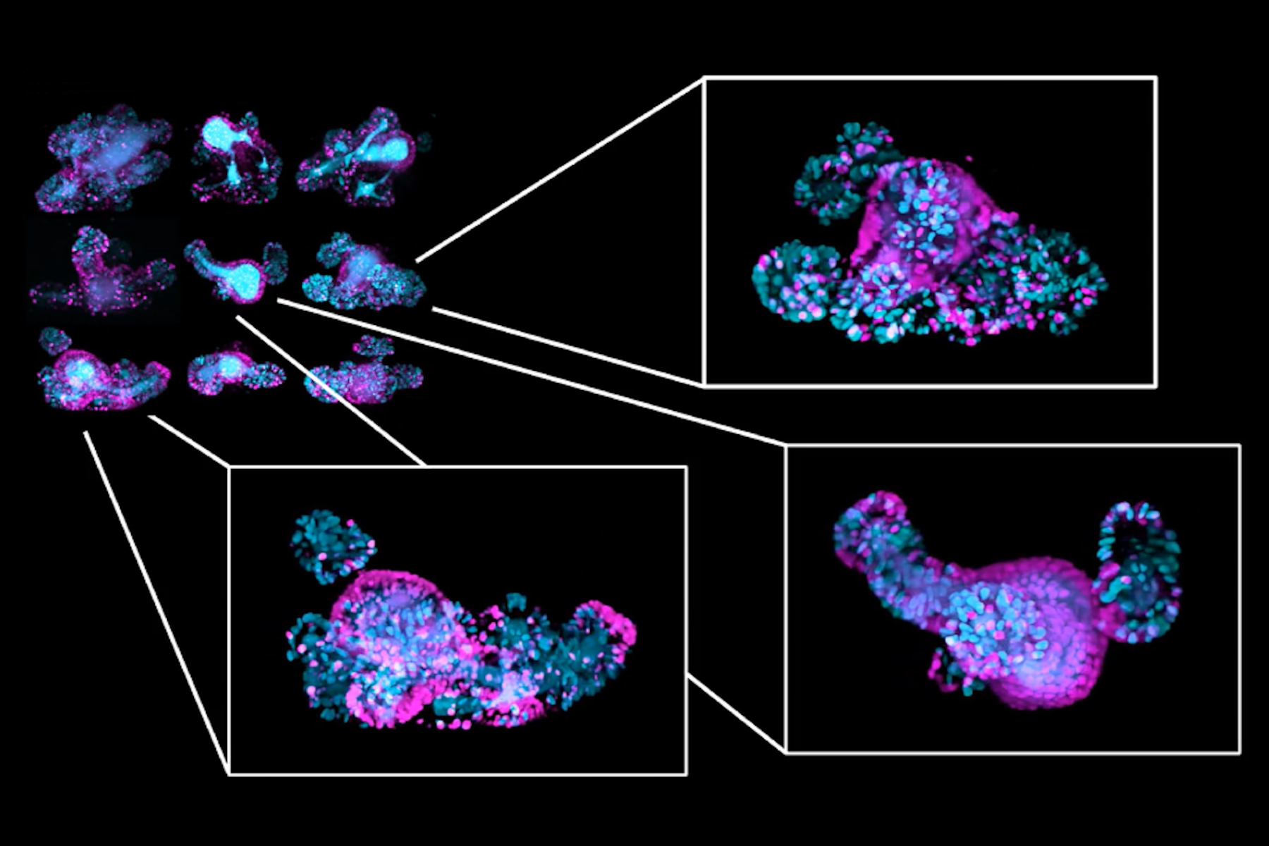
First, Dr. Gustavo Quintas Glasner de Medeiros walks through high-throughput fixed time courses: dissociate to single cells, plate multi-well plates, fix plates at staged timepoints and acquire 3D datasets at scale on a high-content imager (spinning-disc confocal is discussed). He explains how this approach supports phenotypic landscape profiling from large numbers of organoids, and how multiplexed immunostaining can add functional readouts by iteratively eluting and re-staining for additional markers.
To conclude the webinar, Dr. Andrea Boni presents the key features of the Viventis Deep Dual View Light Sheet Microscope and introduces its new imaging option for cleared samples. He explains how the Viventis Deep can be used for 3D live imaging and for cleared sample imagaing simply by exchanging the objective block.
His examples shown include cleared organoid-scale samples up to larger mouse specimens, such as a cleared gastroloid imaged in three colors at single-cell resolution and a tiled mouse salivary gland explant dataset.
Key learnings
- High-content fixed timepoints for statistical confidence: fixed time courses at scale are positioned as essential for generating enough statistical data for phenotypic landscape profiling and classification across variability.
- Light sheet live imaging for dynamics and causality: light sheet microscopy is presented as tunable for organoid imaging and suited to long-term live acquisition where phototoxicity is a constraint.
- Multi-view strategies to compensate for scattering: multi-view illumination/detection and fusion are shown as a route to improve volumetric information deeper in tissue compared with single-view limitations.
- Data extraction that supports discovery: tracking, segmentation, feature extraction, and lineage-tree visualization are used to explore live imaging data and connect dynamics to measurable features.
- Standardization and collaboration to process at scale: the webinar emphasizes shared processing standards and reproducible workflows.
Related Articles
-
 and phalloidin (magenta), imaged using Viventis SCAPE; scale bar 50μm. Courtesy of Marina Cuenca and Heleen Jungen (Dayton lab), EMBL Barcelona.")
What’s the Best Organoid Imaging Approach for Early Drug Discovery?
Organoids and other complex in vitro models (CIVMs) are becoming increasingly important in early…
Jun 30, 2026Read article -
 2D slices of a 1 mm diameter midbrain neural organoid stained with DAPI (blue, nuclear stain), β-tubulin (green, neuronal stain), and GFAP (red, astrocyte stain).")
Fast, High-Contrast Widefield Imaging of Optically Challenging Samples
Live‑cell imaging of large, complex biological samples often requires large fields of view,…
Jun 25, 2026Read article -

History, Developments and Trends of Microscopy in Cancer Research
Cancer is a global disease, with 18 million new cases diagnosed and 10 million cancer-related deaths…
Mar 16, 2026Read article
