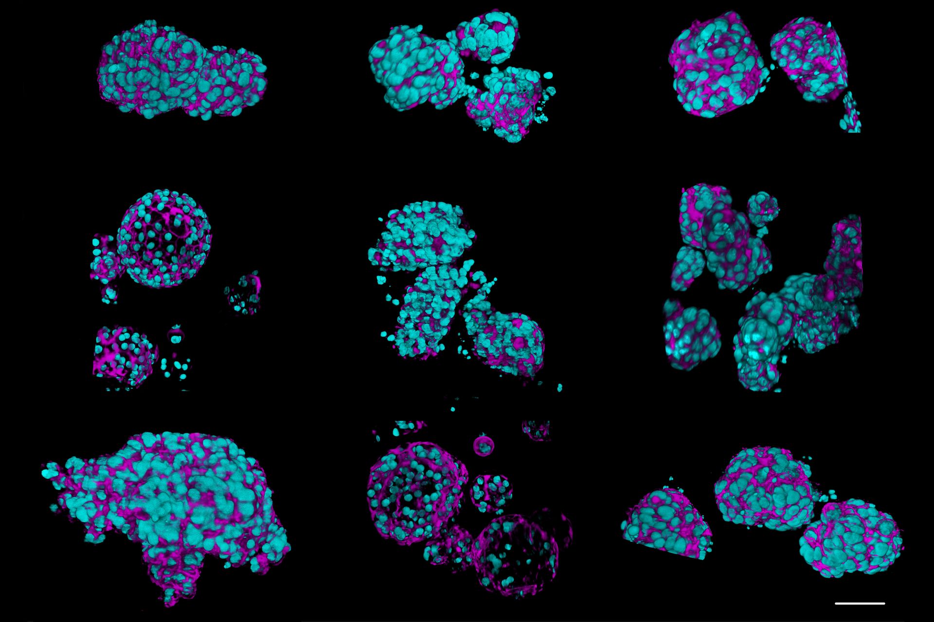
 and phalloidin (magenta), imaged using Viventis SCAPE; scale bar 50μm. Courtesy of Marina Cuenca and Heleen Jungen (Dayton lab), EMBL Barcelona.")
Introduction
Organoids and other complex in vitro models (CIVMs) are becoming increasingly important in early drug discovery and translational research, driven by the need for more predictive, human-relevant data in line with evolving regulatory expectations. However, scientists face many challenges when imaging and analyzing these 3D systems. This eBook explores how complementary imaging and analysis workflows can help you extract deeper insights from complex 3D biological models.
What’s new in the second edition of this eBook?
The updated eBook includes additional real-life case studies and expands significantly on next-generation imaging approaches that can capture meaningful biological data.
- Light sheet imaging workflows: Gentle, high-speed 3D imaging for thick and dynamic samples
- SCAPE technology (single-objective light sheet): High-throughput imaging directly in multi-well plates
- Cleared sample imaging: Visualization of intact structures deep within large organoids
- Long-term live imaging: Tracking cell behavior, interactions, and development over time
If you’re asking these questions:
- How do I image deep into organoids gently over time?
- How can I image cleared organoids?
- How do I scale organoid experiments?
Download the eBook to help find the answers.
Why organoid imaging matters for drug discovery and translational research
Why are traditional models no longer enough?
Traditional models such as animal studies and 2D cell cultures often fall short in predicting human responses, driving demand for more physiologically relevant systems. Organoids and other complex in vitro models are increasingly adopted to address this gap, supported by scientific and technological advances.
What can organoid imaging enable?
Organoid imaging enables you to:
- Visualize cellular interactions and tissue architecture in 3D
- Track development, disease progression, and treatment response over time
- Improve predictive power in early drug discovery workflows
These models replicate key aspects of in vivo biology, offering more relevant insights than conventional approaches and supporting faster, better-informed decisions.
Why is this critical now?
The shift toward new approach methodologies (NAMs), driven by evolving regulatory requirements such as the FDA’s Modernization Act 2.0 is increasing demand for human-relevant models, as researchers aim to:
- Shorten experimental cycles
- Improve translational relevance and reduce attrition rate in drug discovery
- Study complex disease mechanisms more effectively
- Move to more ethical and cost-effective models
What is the best imaging approach for organoids?
There is no single “best” approach—different techniques answer different biological questions. The optimal imaging strategy depends on your experimental goal—whether you need high-resolution structural characterization, long-term live imaging, or fast higher-throughput screening. Rather than limiting yourself to a single technique, combining complementary approaches within a workflow often delivers the most complete insights.
.")
Case studies solving real-life challenges
The eBook highlights how advanced organoid imaging workflows are helping to solve real research challenges, including:
High-throughput airway organoid characterization
- Compare healthy vs. tumor-derived organoids at scale
- Capture 3D architecture across hundreds of samples
Advancing uterine regeneration therapies
- Acquire high-resolution images of whole long sections of uterine organoids, at high-speed
- Reduce imaging complexity while increasing data throughput
Spatiotemporal tracking of intestinal organoid development
- Track cell lineage and development over time
- Combine live imaging with immunostaining to extract additional functional insights
End-to-end organoid imaging workflows
The connected workflow approaches outlined in this eBook support different stages of organoid research:
- 2D pre-culture monitoring and 3D expansion checks
- High-speed organoid throughput imaging
- Long-term live and cleared organoid imaging
- High-resolution, multi-modal imaging with techniques such as CRS and FLIM
- 2D/3D AI-based image analysis
Find the right imaging approach for your organoid workflow.
 2D slices of a 1 mm diameter midbrain neural organoid stained with DAPI (blue, nuclear stain), β-tubulin (green, neuronal stain), and GFAP (red, astrocyte stain).")


